Three-dimensional scans of two mummified newborn woolly mammoths recovered from the Siberian Arctic are revealing previously inaccessible details about the early development of prehistoric proboscideans. The research, conducted in part by American Museum of Natural History Richard Gilder Graduate School student Zachary T. Calamari, also suggest that both animals died from suffocation after inhaling mud. The findings were published July 8 in a special issue of the Journal of Paleontology.
“These two exquisitely preserved baby mammoths are like two snapshots in time,” said Calamari, who began investigating mammoths as an undergraduate at the University of Michigan working with paleontologist Daniel Fisher. “We can use them to understand how factors like location and age influenced the way mammoths grew into the huge adults that captivate us today.”
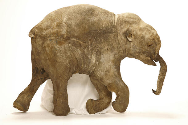
The newborns, Lyuba and Khroma, died at ages one and two months, respectively, and are the most complete and best-preserved baby mammoth specimens ever found. The researchers refer to both calves as mummies due to the high level of soft-tissue preservation, including muscle, fat, connective tissue, organs, and skin. Lyuba’s full-body computed tomography (CT) scan, completed using an industrial scanner at a Ford Motor Company testing facility in Michigan, was the first of its kind for any mammoth.
“This is the first time anyone’s been able to do a comparative study of the skeletal development of two baby mammoths of known age,” said U-M’s Fisher, the lead author of the paper. “This allowed us to document changes that occur as the mammoth body develops. And since they are both essentially complete skeletons, they can be thought of as Rosetta Stones that will help us interpret all the isolated baby mammoth bones that show up at other localities.”
Lyuba and Khroma lived more than 40,000 years ago and belonged to mammoth populations separated by roughly 3,000 miles. Lyuba was found in May 2007 and Khroma was found in October 2008.
The first CT scans of Lyuba were done in Tokyo in 2009 and in Wisconsin in 2010, using medical scanners. But because of Lyuba’s size (about 110 pounds and slightly smaller than a baby elephant), the researchers could not acquire 3D data from her entire body. The researchers finally succeeded in October 2010 at Ford Motor Company’s Nondestructive Evaluation Laboratory in Livonia, Michigan, using a scanner designed for finding flaws in vehicle transmissions.
Khroma’s CT scans were done at two French hospitals. Micro-CT scans of teeth from both mammoth calves were conducted at the University of Michigan School of Dentistry. From the dental studies, Fisher and colleagues determined that Lyuba died 30 to 35 days after birth and estimated that Khroma’s age at death was between 52 and 57 days.
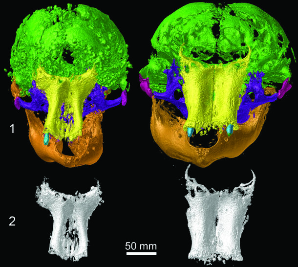
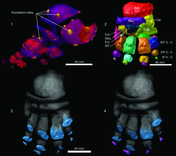
Scans of Khroma’s skull showed she had a brain slightly smaller than that of a newborn elephant, which hints at the possibility of a shorter gestation period for mammoths. Lyuba’s skull is conspicuously narrower than Khroma’s and her upper jawbones are more slender, while Khroma’s shoulder blades and foot bones are more developed. These differences may simply reflect the one-month age difference between the calves, or they could relate to the different populations from which the two calves derived.
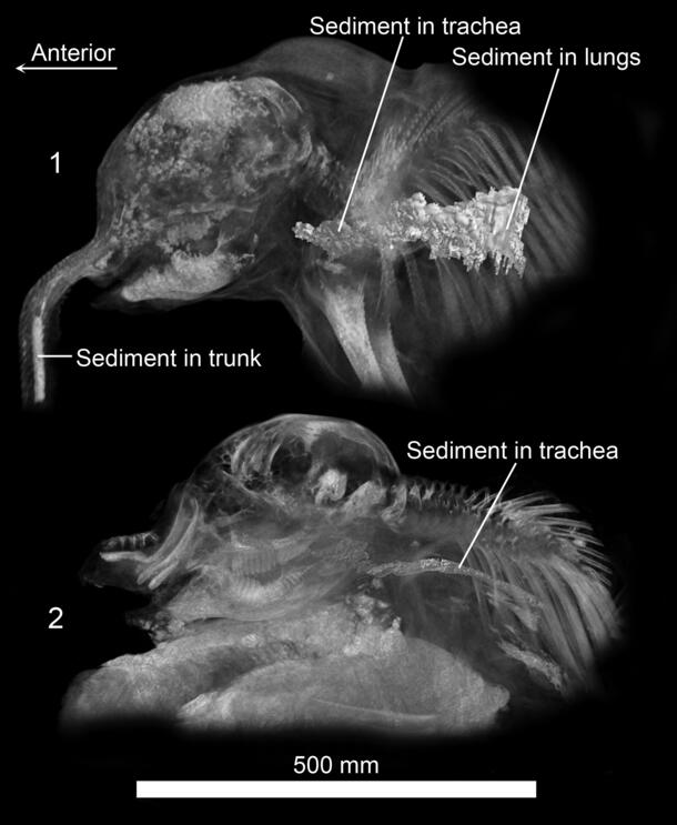
In addition to providing unprecedented insights into mammoth development, the CT scans of Lyuba and Khroma suggest that both youngsters died after inhaling mud, then suffocating. This death scenario was raised for Lyuba shortly after she was first examined. The Khroma CT scans demonstrate that she may have suffered a similar fate.
In Lyuba, the scans revealed a solid mass of fine-grained sediment blocking the air passages in the middle of the trunk. Sediment was also seen in Lyuba’s throat and bronchial passages. If Lyuba had died by drowning rather than suffocation—as some have suggested—then traces of sediment should also have been detected in parts of the lungs beyond the bronchial passages, but that was not the case.
Slightly coarser sediment was found in Khroma’s trunk, mouth, and throat. The animal’s lungs weren’t available for study because they were scavenged before the carcass was recovered. Since both animals appear to have been healthy at the time of death, a “traumatic demise” involving the inhalation of mud and suffocation appears to be the most likely cause of death in both cases, according to the authors.
The researchers suspect that Lyuba died in a lake since sediments found in her respiratory tract include fine-grained vivianite, a deep blue iron- and phosphate-bearing mineral that commonly forms in cold, oxygen-poor settings such as lake bottoms.
A possible death scenario for Khroma places the calf and her mother on a riverbank in the spring. Khroma had been nursing less than an hour before her death, as evidenced by undigested milk found in her stomach during a necropsy by a team of scientists that included Fisher. Perhaps the riverbank collapsed and the two mammoths, mother and daughter, plunged into the river. A fall would account for the fractured spinal column revealed by Khroma’s CT scan, as well as the mud she inhaled.
For more details on this work, see the Museum’s press release.