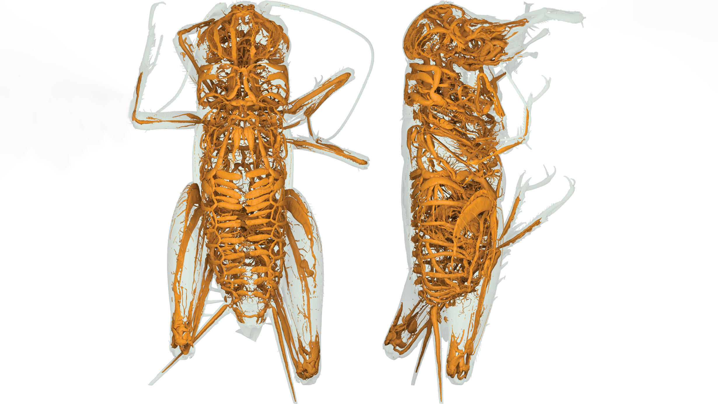
 Three-dimensional high-definition micro-CT scan of a field cricket’s respiratory system shows this species’ unique tracheal architecture.
© Hollister Herhold
Three-dimensional high-definition micro-CT scan of a field cricket’s respiratory system shows this species’ unique tracheal architecture.Insects have long been known to rely on a network of fine tubules, called trachea, that carry oxygen and carbon dioxide directly to and from tissues.
But peering inside this respiratory system—intricate and laborious to dissect—has long been a challenge. Now, a new study published this month in the Bulletin of the American Museum of Natural History uses micro-CT scanning to reveal the respiratory networks of 29 species of insects, creating a new consistent, three-dimensional visual atlas of insect tracheal systems.
“Previously, the way you explored tracheal architecture was through dissection, distorting the morphology of the living insect,” said the study’s lead author Hollister W. Herhold, a doctoral candidate in comparative biology at the Museum’s Richard Gilder Graduate School. “With micro-CT scanning, we’re able to look at everything in a non-destructive way, in situ, to see where all of these trachea lead through the body of the insect.”
All insects, Earth’s most diverse and abundant animal group, use thin-walled air-filled tubes that carry oxygen and carbon dioxide directly to and from tissues, unlike vertebrates, which have gills and lungs and transport these gases in the bloodstream.
Previous studies of insect trachea relied on dissection and focused on smaller numbers of insects, but this new study – conducted over six years – used high-resolution micro-CT scanning to visualize holistic pictures of the respiratory systems of 13 unique insect orders.
For the study, researchers collected insect species like grasshoppers, mantises, and roaches, most found locally in New York and New Jersey. Using high resolution micro-CT scanning, they identified pathways of air in each specimen, indicating the shape and structure of that species’ respiratory system.
Finally, a dynamic map of each insect’s respiratory system was created in a complex, manual process in which the tracheae were filled in using 3D tracing software. The team then examined differences among respiratory systems and morphological traits, such as flight and thermoregulation.
“This work lays an expansive foundation for future studies on the physiology of insect respiration, flight, thermoregulation, and even sound production and hearing,” said co-author David A. Grimalidi, a curator in the Museum’s Division of Invertebrate Zoology.
The three-dimensional models have been made freely available in open-source formats, which the researchers hope will provide an effective platform for continuing research into insect respiration.
“Aristotle was one of the first to look at insects as a scientist. He figured that if insects didn’t have any blood, they didn’t need to breathe, and that idea stuck for over 1,000 years,” said Herhold. “There are a lot of things people assume about bugs that aren’t true. Our big goal was to make it possible for future scientists to look at things that aren’t studied yet.”
Other authors of this study include Steven R. Davis, postdoctoral fellow in the Division of Invertebrate Zoology and assistant professor at Kanazawa University, Kanazawa, Japan; and Samuel P. Degrey of the Kimberly Research and Extension Center.
Insects, which can be found around the globe in nearly every terrestrial habitat, will be showcased in the new Susan and Peter J. Solomon Family Insectarium, opening in Spring 2023 as part of the Museum’s Richard Gilder Center for Science, Education, and Innovation.