A new study published in Science this week by an international research team, including scientists from the Museum, describes the speedy recovery of the meteorites and reports that this space rock is an unusual example from a rare group known as carbonaceous chondrites, which contain some of the oldest material in the solar system. The study of these meteorites and others like them could hold answers to unsolved mysteries about the origin of life on Earth as they contain molecules such as water and amino acids.
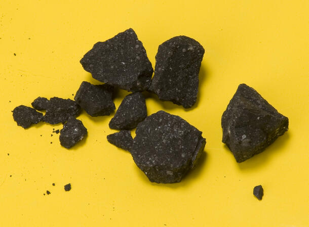
About eight months ago, several Doppler weather radars detected a hail of rocks following a fireball traveling at a record-breaking 28.6 kilometers per second (about 64,000 miles per hour) over the Sierra Nevada in northern California. An immediate search-and-recover mission, led by NASA Ames Research Center, the SETI Institute, and the University of California, Davis, resulted in the retrieval of 77 meteorites. The fragments, which were in pristine shape despite entering the atmosphere at a speed twice as fast as a typical meteorite fall, were collectively called the Sutter’s Mill meteorite after the nearby historical site that started the California Gold Rush.
Several fragments were sent to laboratories around the world for simultaneous analysis of the meteorite’s mineralogy and structure. Denton Ebel, chair of the Division of Physical Sciences at the Museum, received five of the Sutter’s Mill meteorites to study using x-ray computed tomography (CT), an imaging technique that takes pictures of the inside of a specimen without destroying it. The Museum’s scanner takes more than 1,000 x-ray images of the object as it rotates inside of the machine. The data collected from these x-rays are then converted by computers to form a 3-D image of the specimen’s interior, one slice at a time, to understand the components of the meteorite.
This video shows a connected series of virtual slices through one of the Sutter's Mill meteorite fragments.
“In the same way that medical tomography, called CAT scanning, is used to image the interior of the human body, CT scanning in a research laboratory allows us to obtain images of the interiors of solid objects, but with a much higher resolution,” Ebel said. “This is a fundamentally important tool not just for looking at rocks but for curating them and figuring out whether anything interesting is inside.”
CT scans at the Museum, and at the University of California, Davis, revealed that no two Sutter’s Mill meteorites are the same. The meteorites contained angular pieces of different composition and density. This suggests that the surface of the asteroid that spawned the meteorites, their “parent body,” is more complex than previously thought.
For more information, see the Museum’s press release.
Visit the Museum's Arthur Ross Hall of Meteorites to learn more about meteorites.