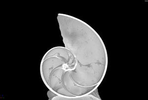
Delicate, with the eerie beauty of a 19th-century engraving, the gray-and-white cross-section of Nautilus pompilius — an object of ongoing research by Museum paleontologist Neil Landman – is the product of a cutting-edge, high resolution, computed tomography (CT) scanner. Acquired by the Museum last summer with a grant from the National Science Foundation, the GE Phoenix V/tome/x Dual-Tube CT Scanner is one of only four of its kind in the country and allows researchers to look deep inside both small and large specimens without destroying them in the process.
“We can see spatial detail not available in dissection, and some parts are so delicate they would be otherwise missed,” says Dr. Landman, curator in the Division of Paleontology who, with geologist Denton Ebel, associate curator in the Division of Physical Sciences, and Curator Darrel Frost, a herpetologist, wrote the successful grant application for the scanner. “Three-dimensional visualization is such an important part of our thinking now—you can put your arms around the object you are studying.”
For each image, the scanner, as a rule, takes 1,500 to 1,700 x-ray images as the sample is rotated in the x-ray beam, at a level of resolution 100 times that of a typical medical scanner used on humans. These images are then used to create a 3D image of the entire specimen—in essence, a stack of virtual dissection slices—that can be manipulated, rotated, and studied from every angle, revealing unprecedented details of its internal structure. “We can only capture so much of the morphology from the surface,” explains Landman. “You want to get insights into the interior.”
In the case of the Nautilus pompilius—a newly hatched specimen recovered in Fiji in the 1930s—Landman is interested in what the interior chambers can tell him about the animal’s buoyancy, a key factor in its survival after birth. Little is known about nautiloids, a group whose ancestors are so old—400 million years—that the extant creatures are called “living fossils.” No one even knows where these invertebrates lay their eggs, which develop slowly to hatch at the largest size of all invertebrates and then take 15 years to reach reproductive maturity. (Landman recently gave a presentation at a Convention on International Trade in Endangered Species of Wild Fauna and Flora [CITES] conference in France, where he cautioned that due to its slow development and rate of growth, “this is an animal that you don’t want to overfish because it may never recover.”)
Since its installation, the dual-tube CT scanner has lent itself to the study of a host of diverse specimens: meteorites by Ebel; rare bat skulls by Nancy Simmons, chair of the Division of Vertebrate Zoology; an early 20th-century knife from Egypt by Alex de Voogt, assistant curator in the Division of Anthropology; the reproductive systems of female spiders by Matthias Burger, a postdoctoral researcher; and the gut contents of a termite entombed in amber by David Grimaldi, curator in the Division of Invertebrate Zoology. The scanner is also accessible to researchers from other institutions, including art conservators who use it to assess fine cracks in antiquities.
Rebecca Rudolph, laboratory manager for the Museum’s Microscopy and Imaging Facility, notes that previously, Museum scientists were forced to go off-site for CT scanning, either to a hospital to use a medical scanner or to a facility such as the University of Texas.
For researchers who intend to cut into a specimen eventually, the CT scanner allows them to zero in on the most promising areas for physical analysis, as well as capture a 3D image of the interior while it’s still intact.
“It takes a lot of guesswork out of the equation,” says Landman.
And those antique engravings and the early naturalists whose discoveries inspired them? Landman said he was thinking only recently about what would happen if someone from another era were suddenly dropped into a 21st-century imaging lab. “They would think it was magic,” he said. “Absolute magic.”
For more about research at the Museum, visit amnh.org/science.
A version of this story originally appeared in the Winter issue of Rotunda, the magazine for Museum Members.