The Museum is home to the largest collection of spiders in the world, one that is still growing through the fieldwork of scientists such as Norman Platnick, Curator Emeritus in the Division of Invertebrate Zoology. Platnick, who has discovered and described more than 1,600 new spider species, says there are many more to find.
“Collections are the only way we can document the plants and animals with which we share this planet,” says Platnick. “For groups that are as poorly known as spiders, there are many areas in the world where they have not been collected at all.”
One of his recent collecting efforts, in late 2010, took Platnick and his team on a month-long expedition into the cloud forests of northeastern Ecuador. Science Bulletins, the Museum’s multimedia program that covers current science, followed the researchers as they worked day and night seeking out spiders from the forest floor to the high canopy.
“Fieldwork is a messy enterprise, which is part of what makes it fun,” explains Platnick. “If you go on an expedition with a purpose that’s too specific, you’re going to come home very unhappy—it isn’t always easy just to go to an area and find one particular species.”
Though Platnick specializes in goblin spiders, a family of tiny arachnids found mostly in forest leaf litter, his international team of arachnologists collects not only every spider they can, but also the insects and other invertebrates they find. At the end of the trip, these samples are packed up and sent to scientists who study those species all over the world.
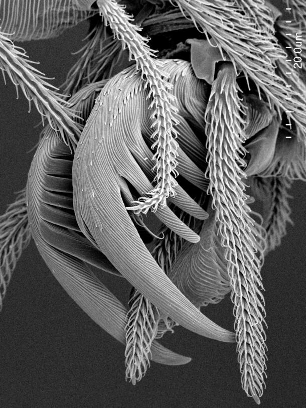
The goblin spiders, however, are sent directly to Platnick and his team in New York. Here, on the Museum’s fifth floor, scientific assistants photograph the microscopic details of each specimen using a high-powered scanning electron microscope (SEM), an immense undertaking.
“Because our spiders are really small, the regular microscope is not powerful enough to show in detail the surface of the spiders,” says scientific assistant Nadine Dupérré. “So the SEM is very useful.” With the SEM, scientists can visualize the individual body parts of each spider, the hairs emerging from between those parts, and even the complex pattern of bumps on the spider’s exoskeleton that may be unique to that species. It’s often the only way to find out whether a particular spider has been seen before or is completely unknown to science.
A version of this story appeared in the the Summer 2012 issue of Rotunda, the Member magazine.