Ventral Trilobite Preservation
We can vividly recall being part of a trilobite hunting expedition that took place in the eastern United States a number of years ago. Over the course of a weekend, literally tons of fossil-bearing rock was excavated by a frenzied group of trilobite enthusiasts, each of whom seemed focused of finding the ultimate Paleozoic prize -- a perfectly articulated dorsal specimen. Utilizing nothing more than crowbars and sledgehammers, and occasionally their bare hands, these dedicated diggers were intent on their task… moving layer after layer of Silurian-age rock. The rhythmic sound of metal banging against stone could be heard echoing throughout the quarry as large chunks of sedimentary matrix began flying this way and that… until the quarry foreman stopped the proceedings with a rather gruff salutation.
As he raised one particular rock in the air, he asked, “who tossed this?” in a voice that could be heard in the neighboring county. “I did,” came a rather sheepish reply from one of the diggers. “I didn't think we were keeping negatives.” Those words served to stoke the quarry boss' anger to the next level. “Negative?” he shouted. “That's not a negative… that's a ventral trilobite! That's what you're all looking for. All you'd need to do is flip it during preparation, and you'd end up with an incredible specimen… but you don't deserve it!”
Whether or not our poor digger deserved to keep his foolishly tossed sample, the fact is that when it comes to trilobites, it's relatively easy to confuse a ventral specimen with a “negative” example. They may initially look the same, but under closer inspection the differences between the two quickly become apparent. Negative, or counterpart, trilobites are exactly that… the reverse impression in the rock caused by the actual trilobite. Often these fossils are left in the field by overzealous collectors who believe that they are of little use, or value to those looking for “true” trilobite specimens.
In contrast, ventral trilobites are complete examples of the actual fossil that have been preserved and subsequently prepared from the reverse, or bottom side. It has been speculated by some scientists that trilobites, much like modern horseshoe crabs, actually swam upside down. Thus the discovery of various specimens “flipped” within the sedimentary bedding plane isn't particularly surprising. When carefully prepared, these ventral examples can often showcase internal muscle attachment “hooks” and scars along with occasional appendage remnants. Many ventral trilobites also display the distinctly pronged hypostome, or mouth plate. These calcified, occasionally forked extensions (which possibly allowed the trilobite to attach themselves to a food source) come in a wide variety of shapes and sizes, and when found independently can often help identify the trilobite species from which they originally came.
Some locations, such as the famed Burgess Shale in British Columbia, the Hunsruck Slate of Germany and the Lorraine Shale of upstate New York, have become renowned for producing trilobites where ventral examples often provide detailed evidence of soft-body part preservation, including antennae, claws, gills and even, on rare occasion, eggs. These specimens are not only prized by collectors around the globe, but are also the subject of intense study by scientists trying to learn more about the often secretive lifestyles of everyone's favorite ancient arthropod, the trilobite.
Here is a look at some ventral trilobites from around the world:
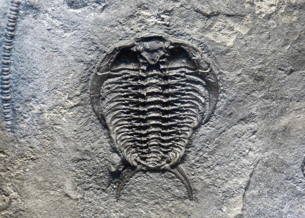
Ceraurus plattenensis (Ordovician, Ontario) is often found ventrally, but is usually "flipped" dorsally for preparation.
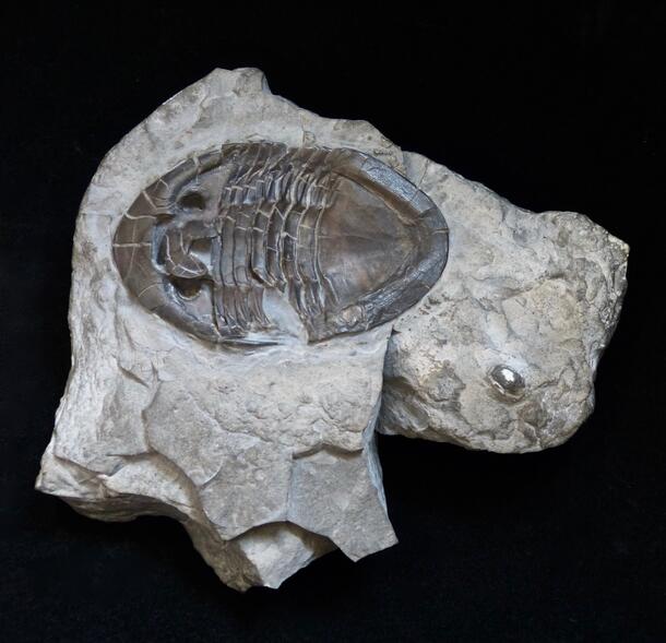
A pair of ventral Asaphus kowalewski from the Ordovician of Russia display the prominent forked hypostome.
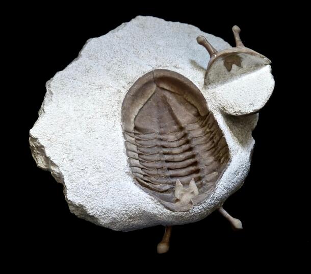
Isotelus mafritzi from the Ordovician of Ontario shows excellent ventral preservation, including the hypostome.
A dramatic Drotops megalomanicus from the Devonian of Morocco. You can see muscle “hook” attachments.
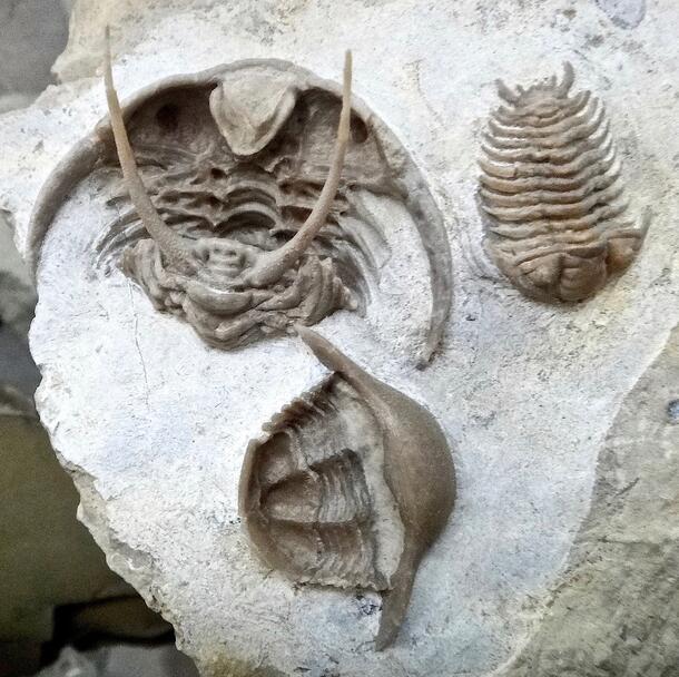
A ventral (and partially enrolled) Gabriceraurus mifflinensis from the Ordovician of Iowa. This trilobite is seen in an association with a ventral Thaleops ovata and a dorsal Ceraurinus sp.
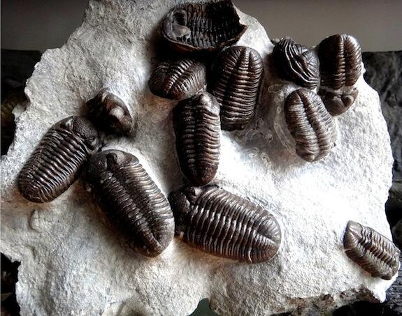
Amid a group of dorsal examples, lurks a single ventrally preserved Eldredgeops milleri from Michigan.
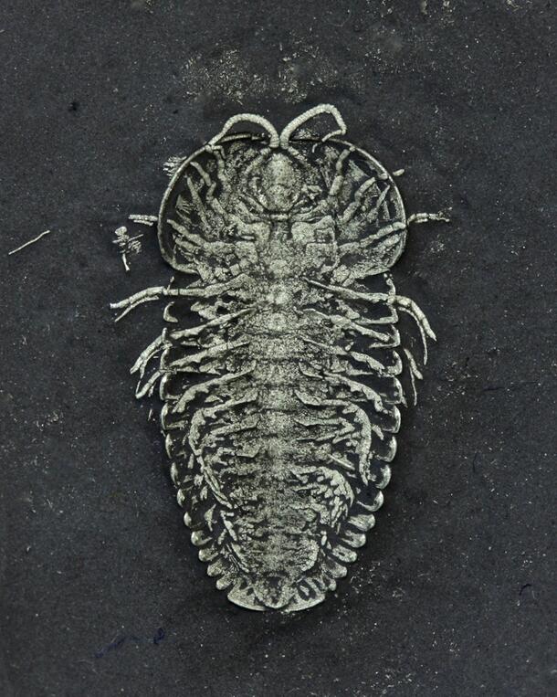
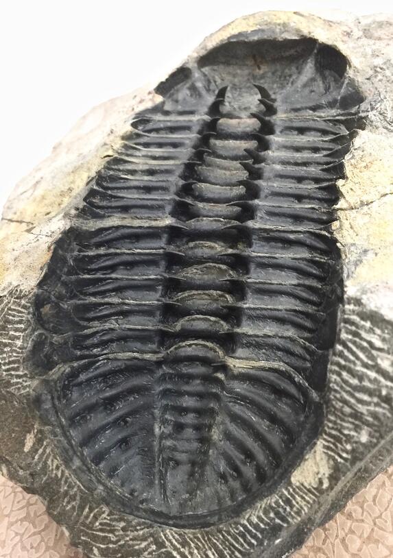
An amazing ventral Triarthrus eatoni from the Ordovician of New York, showing legs and gills.
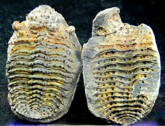
A Bouleia dangicourti from the Devonian of Bolivia, showing a true positive-negative split. There is no element of the actual trilobite in this counterpart.