The latest episode of Shelf Life introduces viewers to foraminifera, microscopic marine organisms known informally as forams.
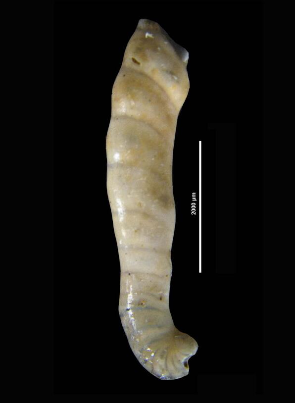
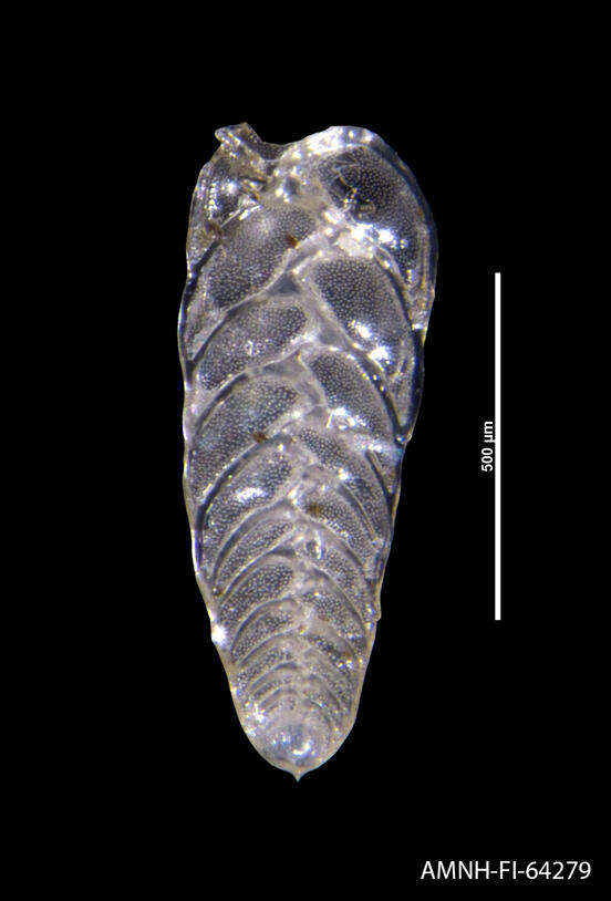
Although foraminifera are single-celled creatures, they have tiny shells that come in an amazing array of shapes. These include spheres like the shell of Globigerina bulloides, above, and simple tubes like that of Hemirobulina gradis, below.
Forams are usually tiny, about half a millimeter long, but larger specimens can measure up to 20 centimeters—about the length of a guinea pig. (Yes, you read that correctly—a single-celled organism the length of a guinea pig.)
Forams have a long and rich history in the annals of science. In the 5th century BC, Herodotus mentioned fossilized foram shells, known as nummulites, which were present in the limestone walls of Egypt’s pyramids. These common fossils were sometimes used as currency in ancient Egypt, and their name comes from the Latin word nummulus, which means “small coin.”
Though there are more than 10,000 recognized species of forams, these organisms can be broken down into two main groups: benthic forams, which live in deep ocean habitats, and planktonic forams, which live in warmer waters closer to the surface.
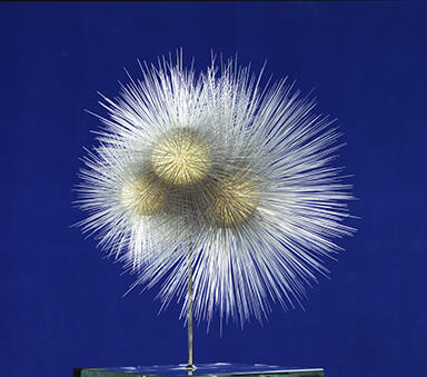
The diversity of forams is reflected not just in their fantastic forms, but in their eating habits as well. Some are predators, eating whatever microscopic organisms—including other forams—they can catch, while others get their meals by filtering tiny pieces of debris out of the water. Some species, like the deep-dwelling Notodendrodes antarctikos, form a system of pseudopods resembling the roots of a tree and sift through the ocean floor looking for food.
Foram shells, known as “tests,” are often composed of calcium carbonate, which means that unlike many other microscopic organisms, they are well represented in the fossil record. Because forams are both abundant and distributed throughout the world, preserved species serve as important indicator fossils, used to identify geologic periods or events like mass extinctions. Researchers can even determine the depth at which other marine fossils would have been found by analyzing the kinds of foram fossils found alongside them—and glean information about ancient climates by analyzing the composition of fossilized foram shells from that time.
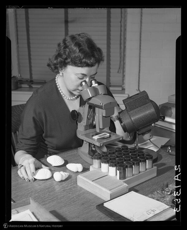
Museum researchers have been contributing to the study of foraminifera for decades. Curator of Micropaleontology Angelina Messina was instrumental in classifying thousands of species of forams in the Museum’s extensive collection. In the course of her work, Messina co-authored a 69-volume Catalogue of Foraminifera with fellow Museum Curator Brooks Ellis and co-founded the journal Micropaleontology.
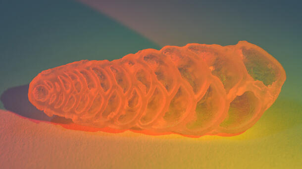
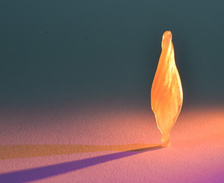
Today, Museum staff are working to digitize the Museum’s extensive collection of microfossils. In addition to creating an online version of the collection that is accessible to researchers around the world, a team of staff members, interns, and volunteers are CT scanning some 50 foram fossils. CT scanning these shells not only makes it easier to visualize them on a screen, but allows for new representations, like 3D-printed models pictured here.