While advances in imaging technologies have opened new pathways for scientists to study natural phenomena, researchers continue to make remarkable discoveries using techniques that have been around for decades. John Sparks, associate curator in the Museum’s Department of Ichthyology, uses enzymes and dyes to reveal key anatomical structures in different species of fishes for study of their function and evolution.
Among his study subjects are ponyfishes (family Leiognathidae), a group of bioluminescent fishes common in the Indian Ocean and Western Pacific that have a light organ. This internal structure, which varies among ponyfish species, surrounds the esophagus and contains luminescent bacteria, the source of the fish’s light. The light organ is larger in males, which have a second species-specific anatomical feature: translucent skin patches, which allow them to use the light organ in displays to attract mates in turbid waters. (Bioluminescent organisms will be explored in the exciting new exhibition Creatures of Light: Nature’s Bioluminescence, which opens at the Museum on March 31, 2012.)
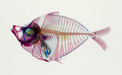
In expeditions to Madagascar, Taiwan, South and Southeast Asia, and other sites, Sparks and his colleagues have collected thousands of specimens, including the Secutor ruconius pictured above. Back in the laboratory, Sparks treats the fish with a series of chemical dyes: red for tinting bones, blue for cartilage, and enzymes to make tissues transparent. Photos of the treated fish—including those featured in the exhibition Picturing Science: Museum Scientists and Imaging Technologies—show the whole body with inner organs intact in striking multicolored images that illuminate structural differences.
Sparks is using this technique, in combination with 3-D imaging, DNA analysis, and other methods, to compare the light organs of different ponyfish species and gain insight into the evolution of light-signaling systems, the role of sexual selection in ponyfish diversification, and co-evolution of the fish and the luminous bacteria abundant in its light organ.
Picturing Science: Museum Scientists and Imaging Technologies is on view in the Akeley Gallery.
This exhibition is made possible by the generosity of the Arthur Ross Foundation.
This story originally appeared in the Fall issue of Rotunda, the Member magazine.