What happens in the brain of a baseball player as a 100-mph pitch hurtles toward him? How does a surfer on her board analyze the nuances of an evolving wave? In a 2013 course for adults, Neuroscience of Sports: Your Brain in Action, experts including Columbia University Medical Center neurosurgeon and neuroscientist Sameer Sheth explained more about the neuroscience of athletics. Dr. Sheth spoke with us about his research into rapid decision-making.

How did you become interested in neuroscience?
I have always had a fascination with the brain and how relatively little we know about it. Although its gross substance resembles a bowl of wobbling Jell-O, its workings are amazingly intricate! How does this bowl of Jell-O enable us to think these incredible thoughts and dreams, interact with people, communicate, and build civilizations? For me, it is a wonderful opportunity to be able to stand back and think about all that.
Could you describe your research at Columbia University?
Clinically, I specialize in functional neurosurgery. I deal with disorders of brain function, like Parkinson’s disease and epilepsy.
My research focuses on decision-making, cognition, and other higher-level brain functions—as opposed to basic motor or sensory functions. As a neurosurgeon I have the ability to use techniques that are somewhat unique, such as electrode recordings of the human brain in action. They allow me to study aspects of the brain that differentiate us from our nearest evolutionary neighbors, non-human primates. Specifically, I study how the human brain has developed to optimize rapid decision-making based on changing cues in the environment.
What will you be discussing in your lecture at the Sackler Brain Bench – Neuroscience of Sports: Your Brain in Action course on Monday, September 30?
I’ll be focusing on one of my particular interests: how the brain is able to rapidly make decisions. This is very applicable to professional athletes. The human brain—especially the pre-frontal region—has developed special mechanisms that allow it to efficiently make rapid decisions by controlling the way it receives and processes cues from the environment.
For example, a professional baseball player engages this mechanism when he is facing a major league pitcher; it allows him to gather an amazing amount of information about the ball’s movement, velocity, and spin in order to make a rapid decision to slightly adjust his swing. On the other hand, the same player taking practice swings with a pitching machine where the ball is coming at him from the same angle and with the same velocity every time does not need to engage that same type of control mechanism.
How are you able to study the brain and learn about these systems in such great detail?
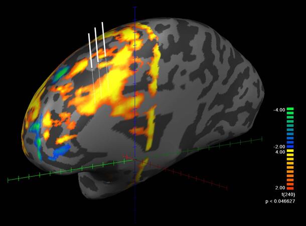
There are a variety of ways to study this. A lot of people have engaged various imaging techniques, like getting an MRI of the brain. There is a particular type of MRI, called functional MRI (fMRI), that allows us to study brain function—not just what the brain looks like, but what parts of the brain are engaged during a specific activity.
The unique ability that we have as neurosurgeons is that we are operating on areas of the brain and in some instances we need to record selective activity in order to successfully preform the surgery. In these cases, if the patient consents, we have the opportunity to study those parts of the brain. During the surgery we can have the patient engage in decision-making tasks such as the Stroop task, which I will describe in my talk, while placing an electrode in a particular part of the brain to record the activity of individual neurons. These types of procedures allow us to study the human brain with a level of precision that is unparalleled.