Museum scientists are getting an up-close look at an extremely rare—and extremely small—shark by taking high-resolution, three-dimensional x-ray scans.
National Oceanic and Atmospheric Administration (NOAA) researchers were trawling in the Gulf of Mexico for a sperm whale feeding study in 2010 when they inadvertently pulled up a tiny, odd-looking shark with a bulbous head and rows of sharp teeth. NOAA researchers subsequently identified the creature as the rare pocket shark (Mollisquama sp.). The specimen is only the second ever collected, 36 years after the first one was found off the coast of Chile.
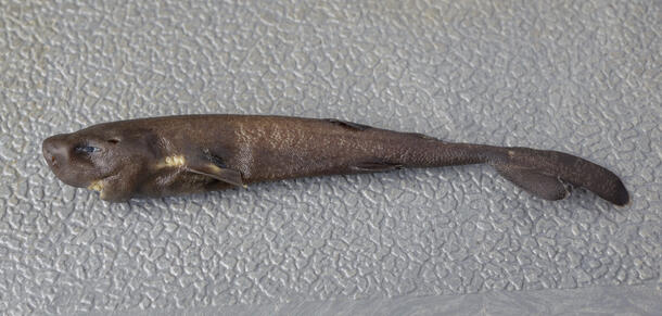
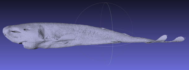
Named for two small openings above its pectoral fins, the pocket shark is still mostly a mystery, as is the purpose its pockets serve. But instead of dissecting this rare 5.5-inch-long specimen, scientists have turned to non-destructive x-ray techniques: computed tomography (CT) scanning in the Museum’s Microscopy and Imaging Facility and at the European Synchrotron Radiation Facility (ESRF) in France.
“The level of detail we can achieve through x-ray imaging is just incredible,” says John Maisey, a curator in the Museum’s Division of Paleontology who has been working with NOAA researchers and Museum Axelrod Postdoctoral Fellow John Denton to scan the specimen. “It allows you to look at these priceless specimens in a way you couldn’t have 10 or 15 years earlier.”

The CT scans proved especially valuable for counting the vertebrae of the pocket shark, the smallest of which were too small to be picked up using standard x-ray imaging, and for counting the teeth. Many of the specimen’s teeth were missing, but by rotating the image of the jaw and examining its inner surface, researchers were able to count the tiny new teeth coming up to take their place.
The species appears to be closely related to cookie cutter sharks, which feed by taking bites out of the skin of larger animals. And the anatomy of the pocket shark’s jaws and teeth indicate that it inhabits a similar ecological niche.
As for the shark’s mysterious pockets, one working hypothesis is that that they might emit a bioluminescent fluid to either attract mates or to confuse predators. Maisey and Denton are now poring over the extremely high-resolution scans taken at ESRF with Mark Grace, the biologist with NOAA’s Southeast Fisheries Science Center who discovered the specimen, to learn more about their anatomy.
But anything gleaned from the scans will likely remain hypothetical until scientists can observe a pocket shark in action.
“I would love to see a pocket shark alive in its environment,” Grace says. “But the CT scans are the next best thing.”