A new study led by Museum scientists provides new insights into a 320 million-year-old shark that was previously only known by its unusual teeth and fragmented jaws. A newly uncovered fossilized brain case of Carcharopsis wortheni, found in the Fayetteville Shale of Arkansas, is helping researchers better place the ancient shark in the tree of life.
Carcharopsis lived during a critical point in evolutionary history, following the end-Devonian extinction event, when nearly 95 percent of vertebrate species went extinct. The late Paleozoic shark was originally described in 1843 based on its distinctive serrated teeth, a feature that is common in modern sharks but rarely found in early shark specimens.

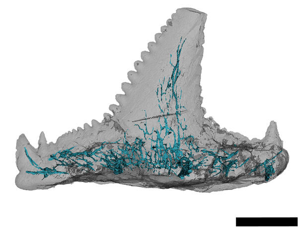
“They look a little like what you’d see in a great white shark, but 320 million years old and with different enamel,” said lead author Allison Bronson, a Ph.D. student in the Museum’s Richard Gilder Graduate School. “This is really early to see serrated teeth.”
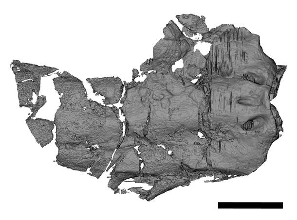
The first known cranium belonging to the extinct shark was discovered in 2007 by Royal Mapes, a retired Ohio University professor and Museum research associate, who donated the specimen along with some 540,000 other fossils to the Museum. Mapes is a coauthor on the new Carcharopsis study, published in the journal Papers in Palaeontology, along with Division of Paleontology Curator John Maisey.
[Watch the video below for more about the Mapes collection at the Museum, including fossil shark specimens.]
The researchers used high-resolution computed tomography (CT) imaging to examine the cranium, a tooth, and an isolated tooth base. Using the scans, they were able to reconstruct the internal canals of the teeth for the first time and found that these are similar to those found in modern sharks.
The arrangement of the shark’s blood vessels—also revealed through CT scans—suggests that Carcharopsis was probably closely related to the group of ancient cartilaginous fish from which today’s sharks and rays evolved. However, more complete fossils are needed to firmly position it in the tree of life.