This instrument is currently broken, and not available for scheduling. E-mail [email protected]if you would like more information. (last updated 03/25/24)
Zeiss EVO 60 Variable Pressure SEMStatusInstrument: GoodEDS Detector: GoodBack Scatter Detector: GoodEBSD Detector: Good
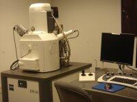
System Overview
The Zeiss EVO 60 is an environmental scanning electron microscope (EP-SEM). In addition to the standard high vacuum (HV) operating mode, the EVO 60 is capable of operating in extended pressure mode (EP). While operating in extended pressure the EVO 60 is capable of viewing specimens that have not undergone the traditional drying and coating preparations. Using small quantities of filtered air and water, the EVO 60 creates a non-destructive coating across the sample preventing charge from building. The EVO 60 also comes equipped with a removable hot/cold stage. When used in combination with the EP mode, the hot/cold stage allows for wet samples to be viewed under the electron beam. Accompanying the EVO 60 are a back scatter detector, energy dispersive x-ray spectrographic detector, and electron back scattered diffraction detector.
EVO 60 Technical Specifications
The resolving power of the EVO 60 is 50 nm and the machine is fitted with a LaB6 crystal filament. The LaB6 provides a brighter image with a higher resolution than commonly used Tungsten filaments. With the benefit of a large sample chamber, the Evo 60 could accomadate samples on the order of size of a basketball (38.5 cm in diameter and 37.5 cm in height).
Backscatter Electron Detector
This system also comes with a removable backscatter electron detector (BSD) in addition to the standard secondary electron detector (SE) used in HV and the variable pressure detector (VPSE) used in EP. The BSD is sensitive to slight energy variations occurring when a high energy electron scatters off the nucleus of an atom. This sensitivity allows the BSD to map out variations in the densities of the sample. On account of the silicone wafer being divided in to four regions allowing it intrinsic investigative properties, the detector type is known as a "Quad."
TEAM™ Pegasus - Integrated EDS and EBSD Analysis
TEAM™ Pegasus incorporates the full range of silicon drift detectors (SDD): the Octane SDD Series with sensors is designed to meet key application needs. Industry leading electronics provide outstanding efficiency and resolution across the full range of count rates.
TEAM™ Pegasus is the answer to your most difficult material characterization problems. By providing both crystallographic and elemental results quickly and easily, TEAM™ Pegasus enables our users to focus their efforts on understanding their materials, rather than on collecting data.
Energy Dispersive X-ray Spectrographic Detectorv
Attached to the EVO60 is an EDAX TEAM™ EDS Analysis System. The EDS detector measures X-rays generated from the sample due to its bombardment from the electron beam. Utilizing this data, the EDAX software is capable of graphing the elemental make up of the sample, as well as creating a color coded map of the sample where different colors pertain to different elements. The Octane Silicon Drift Detector (SDD) can provide a high resolution and accurate map and/or graph of the sample in approximately an hour. Position tagged spectrometry allows for point-specific analysis of all elements from atomic number 4 (beryllium) up to 95 (americium) contained in the sample to be detected and analyzed simultaneously.
Electron Back Scattered Diffraction
The other half of the Pegasus System is the TEAM™ EBSD Analysis System. Accelerated electrons in the primary beam of a scanning electron microscope can be diffracted by atomic layers in crystalline materials. These diffracted electrons can be detected when they impinge on a phosphor screen and generate visible lines, called Kikuchi bands, or "EBSP's" (electron backscatter patterns). These patterns are effectively projections of the geometry of the lattice planes in the crystal, and they give direct information about the crystalline structure and crystallographic orientation of the grain from which they originate. When used in conjunction with the data base that includes crystallographic structure information for phases of interest and with TEAM software for processing the EPSP's and indexing the lines, the data can be used to identify phases based on crystal structure and also to perform fabric analyses on polycrystalline aggregates.