Coated vs. Uncoated Imaging
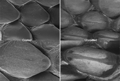
The Evo 60, as previously stated, is capable of imaging uncoated samples. The image above shows the same Bull Snake (Pituophis catenifer) shed before and after it had been coated. Although the images are different, no real detail or resolution is lost. Coated samples can resolve at higher magnifications, but for low and medium magnification the images are essentially the same.
Why are there darker regions in the uncoated images that are not present in the coated images? Samples are coated with a thin layer of gold-palladium or carbon to increase the conductivity. The more conductive your sample is the better image you will be. What an extended pressure scanning electron microscope (EP-SEM) does is emit a small amount of filtered air or water vapor around your sample, decreasing the pressure. The air or water vapor “coats” your sample, increasing conductivity. When the electron beam hits a coated sample it is actually hitting gold-palladium and therefore creates a uniform image. When the electron beam hits an uncoated sample, parts of the sample might not reflect electrons as well as other parts. Hence the difference in coloration.