Examination and Imaging
A variety of examination techniques are used by conservators to observe and document the objects that they care for. These techniques utilize different lighting conditions and levels of magnification, and aid in answering questions about material composition, methods of construction or specimen preparation, impacts of exposure to light and oxygen, pest activity, and past conservation treatment, etc.
Visual Examination
The conservator starts with a general direct visual examination of the object/specimen, sometimes coupled with other senses (such as touch and smell). This examination is usually done in good lighting conditions and sometimes with the aid of low magnification (I.e. an OptiVISOR or magnifying glass). The conservator records her observations in a report using a precise and technical terminology to describe materials and condition issues (such as aging, damage). The documentation generated also relies heavily on imaging, as well as diagrams and annotated images.
From general observations, the conservator may investigate more specific phenomena and hypothesis using more sophisticated visual techniques, as well as analytical techniques. These techniques can provide further details regarding material composition, construction and manufacture, past repairs and mounted methods, physical and chemical decay, etc.
Optical Microscopy
Working under a range of magnifications, either directly observing an object or focused on a small sample collected for study, microscopic examination can yield an extraordinary amount of information about material identification, construction techniques, and condition issues. Stereomicroscopes that offer generous clearance under the lens can be particularly useful. Delicate treatments can also be undertaken under stereomicroscopes.
The Science Conservation lab maintains equipment for standard optical stereomicroscopy, as well and polarized light microscopy (PLM) and cross-section microscopy (CLM).
Digital Photomicroscopy
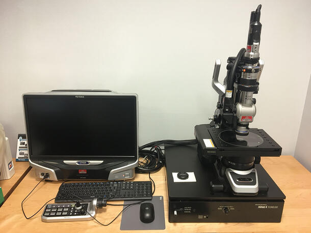
Digital microscopy couples a microscope with a digital camera, providing the ability to capture what you see through the optics of the microscope in still and/or video formats. The camera and microscope may be integrated components of a single piece of equipment, or they may be separate pieces of hardware that have been selected to work together. Digital microscopy offers the chief benefit of facilitating easy capture of photomicrographs. Better component integration, including a desktop computer and monitor, improves the user’s viewing experience and further streamlines image processing, sharing, and archiving.
For mobile and/or low-magnification applications, the Science Conservation lab uses a USB microscope with a gooseneck clamp stand. This tool is cost-effective, straightforward to set up and use, and highly portable. For many purposes it provides the examination and imaging capabilities needed to understand how something is constructed, inform a treatment decision, or identify a trapped insect for IPM. For higher magnification applications, the lab maintains a freestanding Keyence digital microscope with 20-2000x dual objective and 20-200x high performance zoom lenses. The microscope is integrated with a desktop computer used to control settings for lighting and image capture, as well as to perform various types of image analysis functions.
Photography
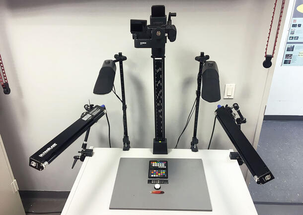
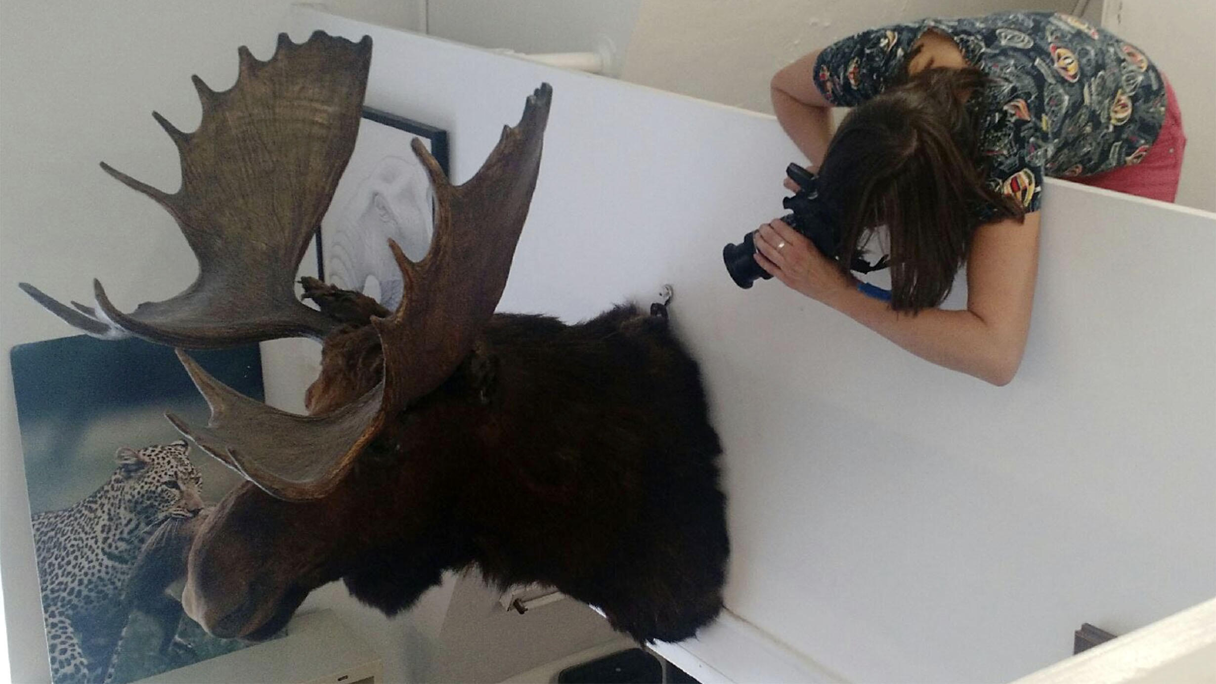
It is part of the conservator’s responsibility to produce photographic documentation of an object’s condition before, during and after its conservation treatment, or as part of survey work as an assessment tool. Conservation imaging follows strict standards and guidelines that are consistent across the conservation field and specialties. It requires standardized workflows for image capture, processing, naming, and archiving. Size scales, color targets, gray scales, and, optimally, light direction indicators, are routinely used. Imaging constitutes a large portion of a conservator’s work and requires constant training as the technology advances fast.
For many decades, best practices for conservation documentation called for Black & White print and color slides, but digital photography is now standard practice. This shift has many advantages in how images can be modified and used, but also raises other concerns about the permanence and preservation of this documentation. Commentary 28 of the AIC Guidelines for Practice includes information on recommended practices in preserving documentation.
The AIC Guide to Digital Photography and Conservation Documentation edited by Warda, Frey, Heller, et.al. is a comprehensive reference intended to maximize the production and preservation of digital images for documentation. It is available in digital and hardcopy formats.
Visible Light Imaging
Several different lighting techniques can be combined with photography to capture images in visible light. Best practice guidelines and basic workflows for visible light photography techniques can be found on the AIC Conservation Wiki.
Multi-Band Imaging
Multi-band imaging (MBI) is a real-time image capture technique designed to supplement what is visible to the unaided eye with the visualization of information in other regions of the electromagnetic spectrum, usually ultraviolet and infrared. MBI requires a modified digital camera, as well as equipment to provide different lighting conditions, filters to control which wavelengths are allowed to reach the camera sensor, and reference targets to standardize exposures and color management.
When examining an object, multiband imaging can reveal features that visibly contrast in the ultraviolet or IR regions, but are otherwise difficult to detect under normal lighting conditions. Those features can provide information about a material’s composition or it’s distribution on a surface, which in turn may offer clues to that material’s origin or purpose. In addition, some materials undergo deterioration processes that can be readily visualized in particular parts of the spectrum. In this way, MBI can be used to document and track changes in an object’s condition over time.