Picturing Science
June 25, 2011 — December 7, 2014

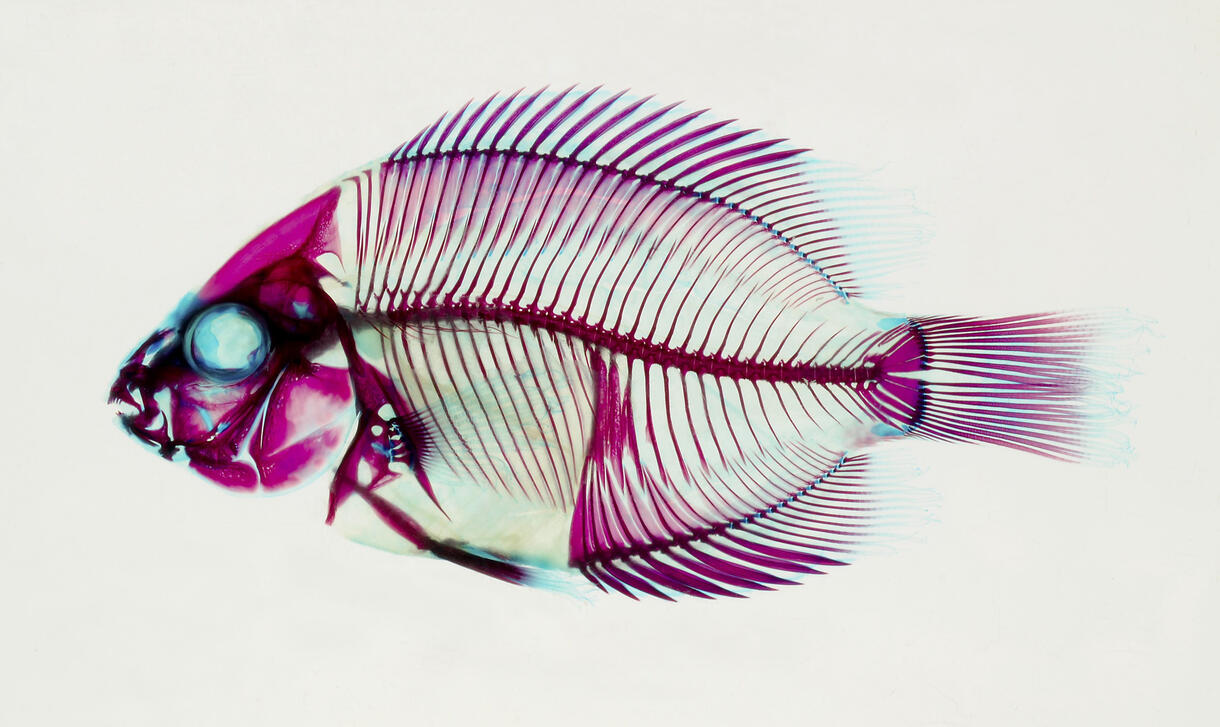
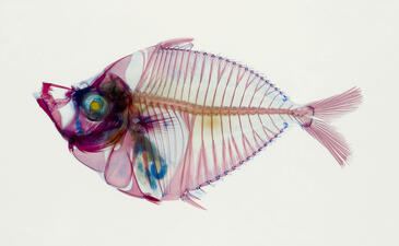
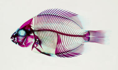
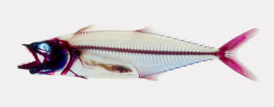
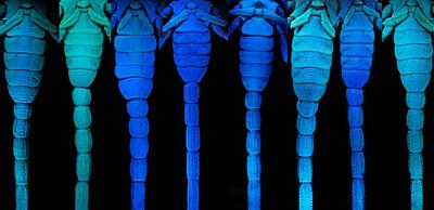
Whether Museum scientists are studying parasites, people, or planets in other solar systems, cutting-edge imaging technologies such as infrared photography, scanning electron microscopes, and CT scanners now make it possible to examine details that were previously unobservable.
This exhibition features more than 20 sets of large-format images that showcase the wide range of research being conducted at the Museum as well as how various optical tools are used in scientific studies.
Biological dyes are used to highlight details of cichlids and other small fish.
J. Sparks/© AMNH
J. Sparks/© AMNH
Biological dyes are used to highlight details of cichlids and other small fish.
J. Sparks/© AMNH
J. Sparks/© AMNH
Associate Curator in the Museum’s Division of Vertebrate Zoology John Sparks, an ichthyologist, highlights details of cichlids and other small fish using biological dyes.
J. Sparks/AMNH
J. Sparks/AMNH
Close-up pictures of insects are not just visually arresting—they also help scientists identify species. Examining the different microscopic features of the many members of the family Vespidae—which includes wasps, yellowjackets and hornets—has been greatly aided by advanced imaging tools. The technique called photomontage brings selected areas of the insect into focus.
J. Carpenter/© AMNH
J. Carpenter/© AMNH
A Museum arachnologist uses UV fluorescence imaging to study scorpions, in this case ten different species of Opistophthalmus.
© AMNH
© AMNH