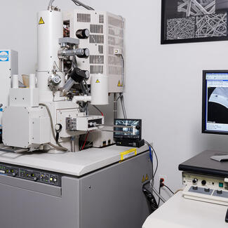
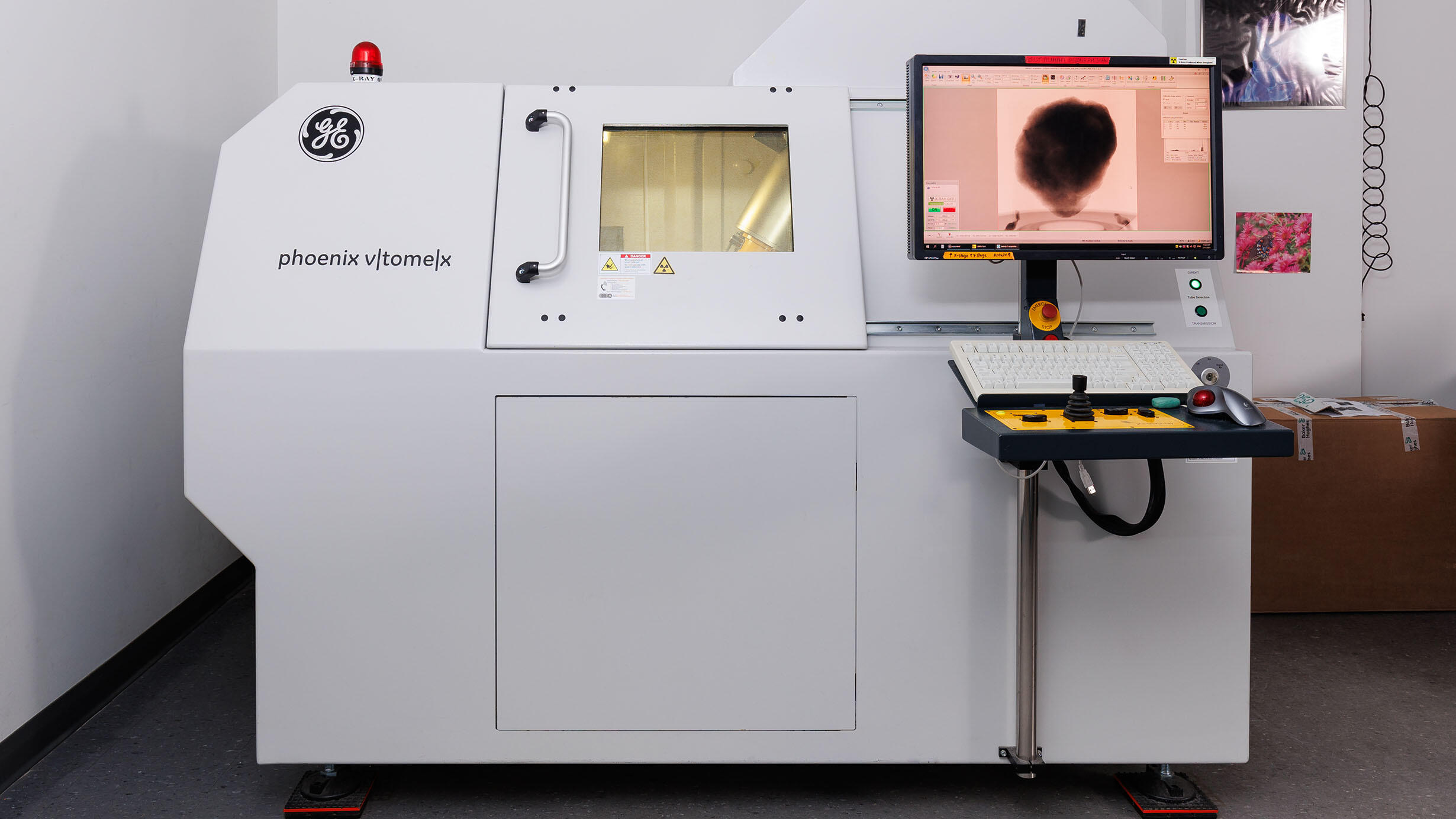
GE phoenix v|tome|x s240
 Daniel Kim/© AMNH
Through capturing of thousands of sequential x-ray images (radiograms) across a 360° rotation of a sample, the CT scanner is able to supply enough information to create a digital representation of the scanned sample's x-ray absorbative material. 3D representations of the physical object can then be investigated and digitally dissected through the use of volume rendering software. These non-destructive methods have enabled the study of internal features of museum collections. This was previously impossible due to the deconstructive nature of other investigative methods.
Our Micro Computed Tomography (μCT) Laboratory has been developed with the intent of doing non-destructive, 3D imaging and analyses of both rare and delicate samples of varying sizes and physical properties. Another aim has been to digitally collect samples for metrology and collaborative purposes due to the ease of file sharing and digital processing.
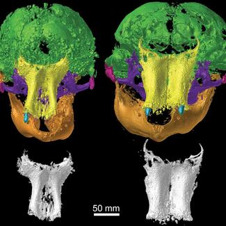
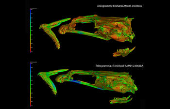
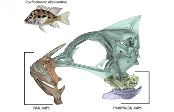
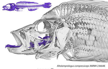
Sample Images: GE Phoenix v|tome|x 240
View Additional Instruments