Instrumental Analysis

In the preservation context, instrumental analysis supports efforts to characterize materials found and used in collections. Analysis may be aimed at identifying adhesives, coatings, paints, etc. used in the production of a specimen or artifact; discerning compounds associated with use, deterioration, pest protection, or repair; or tracking physical or chemical change with time and exposure to the agents of deterioration.
FTIR Spectrometry
Fourier-Transform Infrared Spectroscopy (FTIR) measures the intensity of absorbed vs reflected/transmitted infrared radiation as it interacts with a liquid, solid, or gaseous sample. The IR source emits a beam that may be absorbed by some of the molecules of the sample but transmitted or reflected by others. At each wavelength, the transmitted or reflected infrared is measured by the detector, and then a mathematical algorithm is used to produce a spectrum. Peaks in the spectrum represent functional (-R) groups within the compound analyzed, so the peak pattern is characteristic of that compound. As a consequence, a spectrum from an unknown sample can be compared to known spectra in a reference library, and assuming it is represented in the library, an identification can be made.
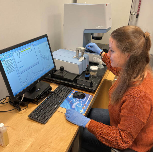
In addition to identifying unknowns, FTIR can be used to observe chemical changes in certain materials. This is done through a series of measurements representing different sample states. For example a sample might be analyzed in roughly the same location over time while it is exposed to light, or before and after treatment with a substance of special interest. Although this approach to analysis is not quantitative, there may be measurable change in peak positions, or in the proportional size of key peaks in the spectrum.
Learn more about how we are using FTIR analysis here.
Spectrophotometry
Spectrophotometry is a non-invasive analytical technique that measures visible light reflected by a material at individual wavelengths across the visible light spectrum. The result is a numerical measurement of color in a sample, which is useful when one needs to describe that color with specificity, and when monitoring color change (i.e. fading) over time. To measure color accurately, a spectrophotometer relies on a light source, a monochromator that isolates and directs the wavelengths, and a photodetector that measures reflected or transmitted radiation.
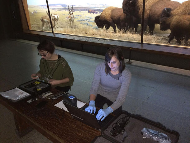
In our lab we have often utilized spectrophotometry in combination with accelerated aging to monitor how samples undergo changes in color with time. Learn more about how we are using spectrophotometry here.
Accelerated Aging Chamber
Accelerated aging chambers are used to rapidly reproduce in samples the damage that is naturally caused by light, temperature, and humidity in real environments over longer periods of time.
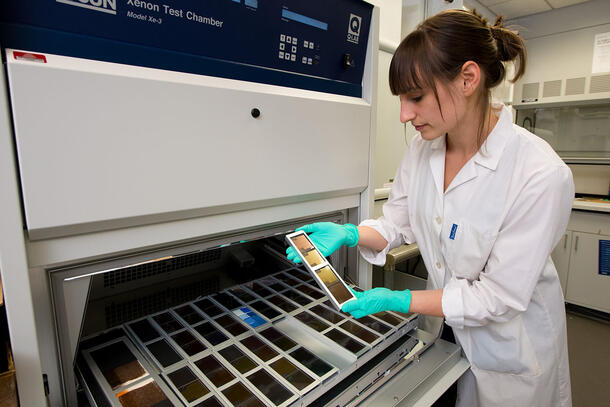
Accelerated aging makes use of the principle that exposure to high intensity light for a short time can produce deterioration similar to that caused by low intensity light over a longer time. However, in order to correctly interpret one’s results, one must understand that for many reasons, accelerated aging does not occur in a way that is strictly reciprocal. In part this is due to the inability of any aging chamber to replicate every aspect of real-world exposures: wet/dry, thermal, or light/dark cycling, the spectrum of incident light, and the presence of air pollutants, dust, or adjacent materials may be impossible to simulate. This non-equivalence is also a consequence of thermal chemistry that unfolds alongside light damage but cannot easily be differentiated from it or accelerated proportionally.
Benchmarking addresses this problem. To create a benchmark, materials aged in real-time are used to define the extent of change taking place over a known duration. When a comparable degree of change is observed in the accelerated test, a correlation factor can be identified to be used in calculating an approximate relationship between accelerated and real-time aging. However, benchmarking has some obvious drawbacks, not the least of which is that a material that ages well may take many years to fail in a real-world exposure environment.
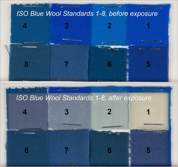
For lightfastness testing, a common benchmarking standard is the Blue Wool scale, eight wool swatches dyed with different dyes that exhibit a range of known lightfastness behaviors. By including the Blue Wool scales in an accelerated light-aging test, one can make a comparison to the sample and provide a rough indication of how quickly it will be expected to fade in real time.
Learn more about how we are using accelerated aging here.