Digital Preparation
 Daniel Kim/© AMNH
Many of the tools and techniques used by today’s preparators have changed little since the origins of the field over 100 years ago.
However, in recent years a number of new technologies that have been applied to preparation are opening up exciting possibilities for paleontological research and preparation.
Three tools of particular utility are:
- High-resolution X-ray computed tomographic (HRXCT) scanning
- 3-D surface scanning
- 3-D printing
The use of these technologies is unlikely to replace traditional preparation methods anytime soon, but for some research projects and for certain specimens they can provide information that would be otherwise unobtainable.
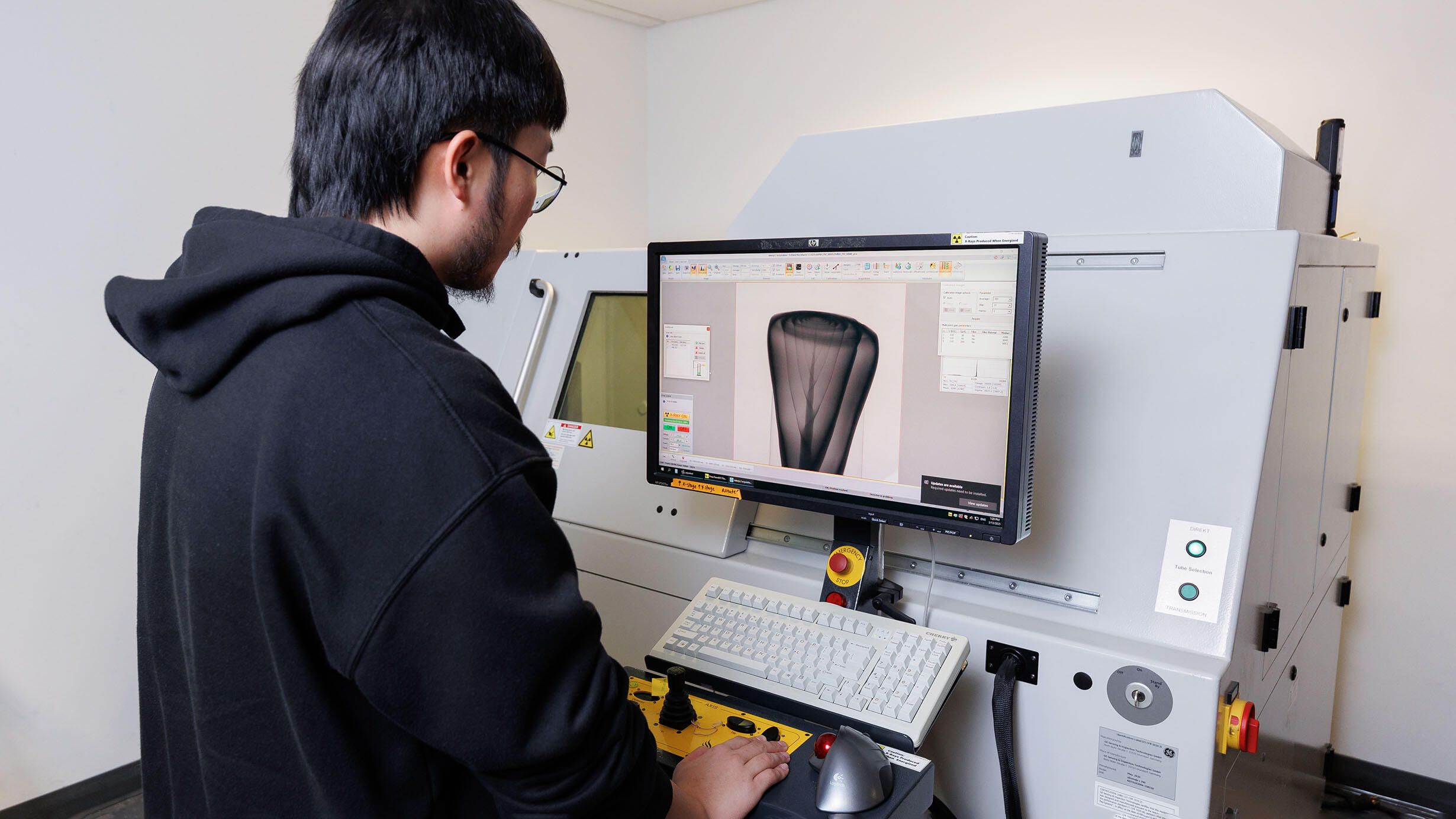
High-resolution x-ray computed tomographic scanning (HRXCT)
HRXCT, also known as X-ray computed micro-tomography (µCT), is a non-destructive imaging technique in which the structure of an object can be visualized by capturing a series of two-dimensional X-ray images as the object rotates between an X-ray source and a detector. The resulting images, or ‘slices,’ are reassembled digitally to create three-dimensional renderings of the object, inside and out.
While medical CT scanners typically employ X-ray energies lower than 140 kV, industrial scanners are capable of generating X-ray energies more than three times as powerful (up to 450 kV or more). This permits penetration of denser materials, like meteorites and fossilized bone, than is possible with medical CT. X-ray microscopy is a specialized application of CT scanning, providing sub-micrometer resolution that allows visualization of bone microstructure. Synchrotron CT uses X-rays generated from a large, circular particle accelerators to scan more quickly, or with better contrast between bone and rock than can be achieved using regular CT scanners.
Reasons to use HRXCT scanning
HRXCT scanning is not appropriate for all types of specimens. It is dependent on having contrast between matrix and bone, which varies greatly. However, in certain applications it can eliminate the need for traditional preparation techniques, thereby potentially saving time and money. Scanning can also aid in specimen conservation as it minimizes the need to handle specimens, both in preparation and in research.
Selecting appropriate specimens
How well a fossil will scan depends on its geometry and its composition. HRXCT scanning geometry is cylindrical, so the more equant a specimen is in the scanning plane, the more scanning artifacts will be minimized. A slab’s geometry is, unfortunately, quite problematic: the X-rays must travel through much more material on one axis than on the other. The composition of the specimen will determine the degree of contrast between the bone and the matrix.
Generally, specimens in a clastic silt or sandstone matrix will scan more successfully than specimens preserved in limestone, because in the latter the calcium phosphate in bone has a similar effect on X-rays as the calcium carbonate in the limestone. Specimens rich in iron do not scan well, because the iron stops a lot of the X-rays. Two specimens from the same locality/formation can scan quite differently due to localized taphonomic regimes. Specimens from some formations, such as the White River, scan particularly well. Again, a quick “scout scan” will indicate whether scanning is a worthwhile investment.
3-D surface scanning
Three dimensional surface scanning is another non-contact, non-destructive technology that can provide information that may be difficult to obtain using traditional preparation techniques. In 3-D laser scanning the specimen is placed on a digitizing platform (often a turntable) and an optically safe laser beam is projected over the surface while cameras record the measurements. The resulting data points are captured by a computer which can then be merged into a 3-D representation of the specimen. An increasingly popular alternative to laser-scanning is structured (or “white”) light scanning, which uses photogrammetry to produce similar surface representations.
3-D scanning is particularly useful when you are only interested in surface detail and when extremely accurate measurements of complicated shapes must be obtained (measurements are can be accurate to ±.0005). Based on a scan, missing portions of a bilaterally symmetrical specimen can be reproduced (e.g. creating a full skull when half remains in the matrix, is lost or distorted). Software also allows for “retro-deformation”, where the scan can be manipulated to allow researchers to correct for deformities in a misshapen specimen.
3-D printing
In 3-D Printing, data files from either a 3-D surface scan or from a High Resolution X-Ray Computed Tomography scan are fed into a rapid-prototyping machine where a three-dimensional model is created, typically by building up layers of thermoplastic resin. These models can often be molded and cast repeatedly if necessary.
The ability to create molds from a 3-D printout means that preparation or modeling of the smallest or most delicate specimens may not be necessary. The technique is particularly useful for extremely small specimens, as the models can be easily enlarged to allow for easier viewing of the smallest morphological features. When the technique uses HRXCT files, internal anatomy can be illustrated by printing; for example, using just one half of a skull, or printing an endocast of the cranial cavity. Data files for the rapid-prototyping machine can be shared digitally allowing others with the technology to create their own cast models, thus increasing research access.
The thermoplastic resins used to create the printed model may not be stable in the long-term, so it is recommended that this original cast be modeled and re-cast in more archival media if longevity and multiple copies are necessary. At this point in the development of the technology the casts created are good, but some resolution is lost and, with the exception of small specimens, a 3-D printed model will not have quite as much detail as a well-made silicone mold.
These Fossil Preparation resources were originally developed in 2007 with the support of the National Science Foundation (NSF).
![]()