Embalmed more than 1,500 years ago, she is one of the most well-preserved mummies from The Field Museum’s foundational collection, 19 of which are on view at the Museum in the special exhibition Mummies.
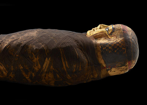
For decades, keeping mummies well preserved also meant severely limiting the ability of researchers to study them. The result is that little was known about the Gilded Lady beyond what could be gleaned from the mummy’s exterior, with its intricate linen bindings, gilded headdress, and painted facial features.
These exterior details do offer some clues. The mummy dates from 30 BC–AD 395, a period when Egypt was a province of the Roman Empire. While the practice of mummification endured in Egypt, it was transformed by Roman influences. Before the Roman era, for example, mummies had been placed in wooden coffins, while the Gilded Lady is preserved in only linen wrappings and cartonnage, a papier-mâché-like material. Also absent are the hieroglyphics that decorated mummy coffins in earlier times.
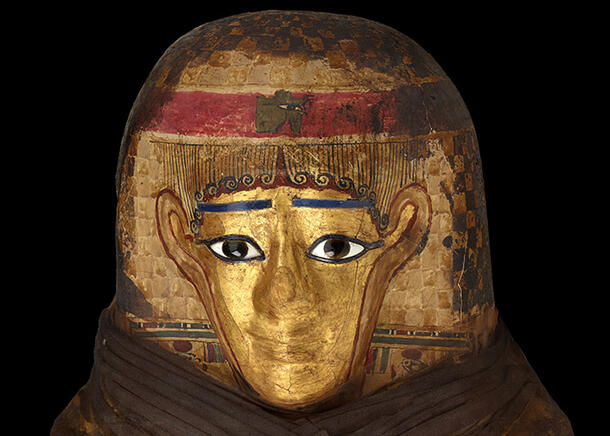
Other traditions surrounding the afterlife persisted, as demonstrated by the Gilded Lady’s intricately painted headdress. Ancient Egyptians believed that in the afterlife, the dead would still require their eyesight, hearing, taste, and smell—and that these senses could be preserved by a mask.
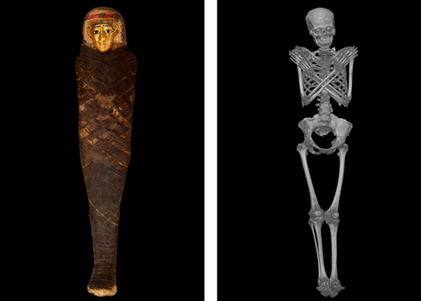
The Gilded Lady’s body case does not reveal much about the woman inside, though. Scientists have long been eager to examine mummies without risking damage to their fragile contents. The first x-rays of a mummy were taken in 1896, just a year after the technology was first developed, points out Museum Curator David Hurst Thomas, who is overseeing Mummies here this spring.
Seeing Inside
Today, computerized tomography (CT) scanning offers researchers glimpses of mummified individuals like never before. By combining thousands of cross-sectioned x-ray images, CT scans let researchers examine the inside of mummies, revealing details about the person’s age, appearance, and health.
“Scans like these are noninvasive, they’re repeatable, and they can be done without damaging the history that we’re trying to understand,” Thomas says.
CT scans like this one of the Gilded Lady reveal that she was probably in her forties. They also suggest that she may have suffered from tuberculosis, a common disease at the time.
Revealing Details
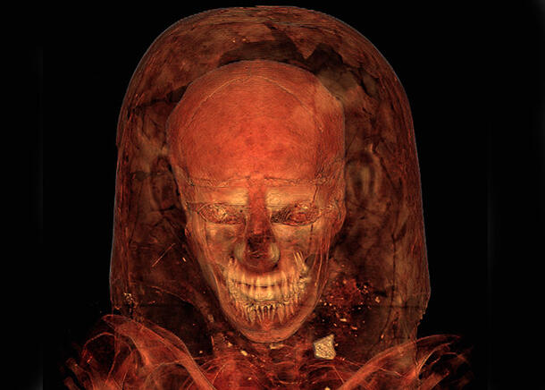
Amazingly, CT scans can also provide details about an individual’s appearance. The Gilded Lady, it turns out, had a slight overbite, as seen in the scan below (top left). Another scan (bottom right) provides one more hint about what she looked like in life: on her scalp, you can see traces of curly hair.
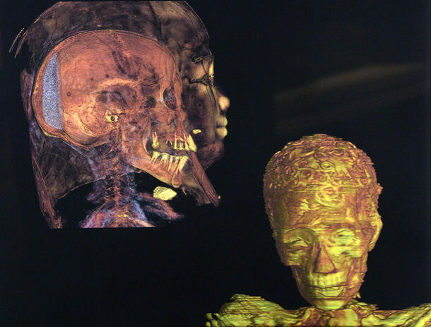
CT scans can also tell us about more than just an individual mummy. The technology helps scientists glean new insights into how ancient Egyptians preserved their dead. In the image above, for instance, the white lumps under the Gilded Lady’s chin and at the back of her skull are thought to be lumps of resin, likely inserted to improve the mummy’s odor.
Face to Face
Using the CT scans of the Gilded Lady, scientists from The Field Museum generated a virtual reconstruction of the skull, then worked with 3D-printing specialists to create an exact physical replica of it without opening or disturbing the actual mummy in any way.
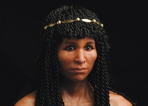
The research team then collaborated with Élisabeth Daynès, an award-winning sculptor noted for hyper-realistic re-creations of fossil hominids including the Australopithecus known as Lucy. Daynès used the detailed skull model, CT scans, and forensic research to create the portrait above: an artist’s interpretation of what the Gilded Lady may have looked like in life.