Shelf Life 11: Green Grow the Salamanders
"We maintain about 80 live cultures of microorganisms, including algae. And about half of them are unique to our collection."
-Eunsoo Kim, Assistant Curator in the Division of Invertebrate Zoology
Collecting The Invisible
The Museum’s collections house many eye-catching items, from the towering Tyrannosaurus to striking totem poles. But there is also a whole category of significant specimens that are imperceptible to the naked eye. We’re talking microbes: single-celled organisms stored in petri dishes or on glass slides.
Microbes at the Museum
This episode of Shelf Life looks at how Museum researchers are currently studying the surprising role single-celled algae play in the life of the spotted salamander—and comes on the heels of the opening of a new exhibition, The Secret World Inside You, that is all about the intricate relationship between humans and the invisible microbes that live on us and in us.
But the Museum first began collecting microbiological specimens and creating exhibitions about the roles of microbes in human health more than a century ago.
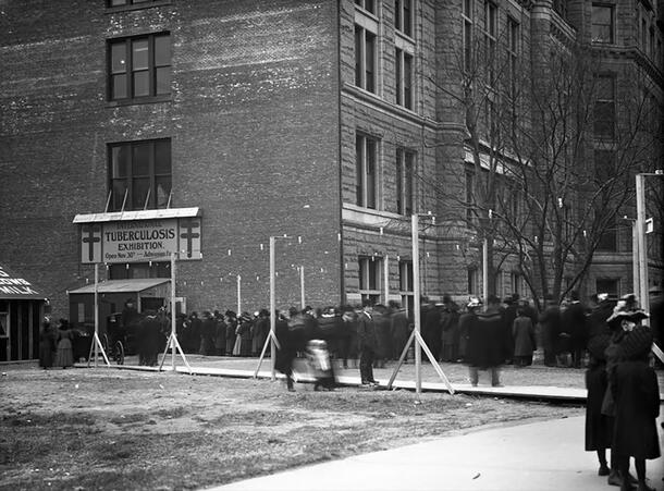
In 1908, the Museum organized an exhibition about tuberculosis, then a common disease and one of the leading causes of death worldwide. Many New Yorkers of the time would have been painfully familiar with the symptoms of the highly contagious illness and its dire consequences.
Far fewer were acquainted with the cause of the deadly disease: Mycobacterium tuberculosis, a strain of infectious, airborne bacteria that attacks the lungs. New Yorkers were so eager for more information about the infection, and about the invisible invaders that caused it, that lines for the Museum’s exhibition snaked around the block. Up to 10,000 visitors saw the show in a single day.
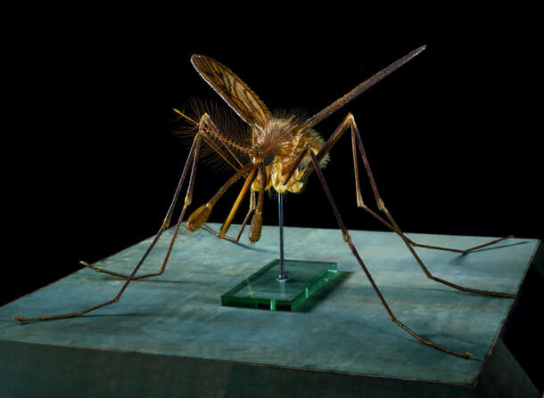
The staggering success of the tuberculosis exhibition spurred the creation of the Museum’s Department of Public Health (DPH), which focused on producing exhibitions and educational materials about the biology behind food safety, water purification, and urban sanitation. One exhibition, developed to educate the public about the spread of diseases, featured a giant model of the malaria-transmitting Anopheles mosquito that’s still on display today.
In addition to public education, the new department’s curator, Charles-Edward Winslow, also pushed for better ways to study the organisms that caused diseases. His efforts led to the creation of a collection of microscopic life, which Winslow called the Living Museum, aimed at helping researchers cultivate, study, and classify different types of microorganisms. Winslow solicited live cultures from all over the country. Within two years, he had gathered 578 bacterial strains and provided 1,700 cultures to researchers at more than 120 labs and universities.
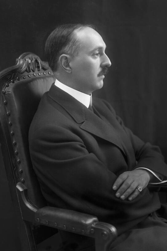
Like other collections, the Living Museum designated type specimens that would serve the same purpose as their counterparts in collections of fishes, insects, or dinosaurs—offering a guiding and definitive specimen against which others could be compared. According to Winslow, it was “the first attempt in this country to standardize these life forms through museum methods.”
A Foundational Collection
The Living Museum—which became known as the Winslow Culture Collection—included some of the most virulent germs around, including bacteria known to cause typhoid, cholera, and, of course, tuberculosis. But it also included beneficial microbes like those used in the production of yogurt, underlining humanity’s complex relationship its microbial neighbors.
When the Department of Public Health was later folded into the Department of Public Education, the Museum’s first-of-its-kind culture collection lived on. In 1925, it moved to a new home at the Army Medical Museum in Washington D.C., where, under the care of the Society of American Bacteriologists (now the American Society of Microbiologists), it became the basis for one of the most important collections of microbial life on the planet: the American Type Culture Collection (ATCC).
Today, when researchers want a sample of an existing microbe species to study, or to describe a new one, they turn to the ATCC. Winslow's library of microbial type specimens has grown and flourished and become the specialized collection he envisioned nearly a century ago, and much more. What began as the Living Museum has become an active and widely used clearinghouse for microbial specimens, cell lines, and more, supplying scientists doing microbial research and medical studies with materials that are central to their work.
Microscopic Marvels
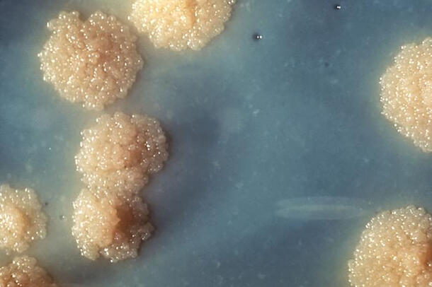
Winslow’s collection of cultures, stored in petri dishes, glass slides, and vials full of glycerol, was the Museum’s first collection of microbes, but not its last. Today, the Museum is home to a number of collections of microscopic life.
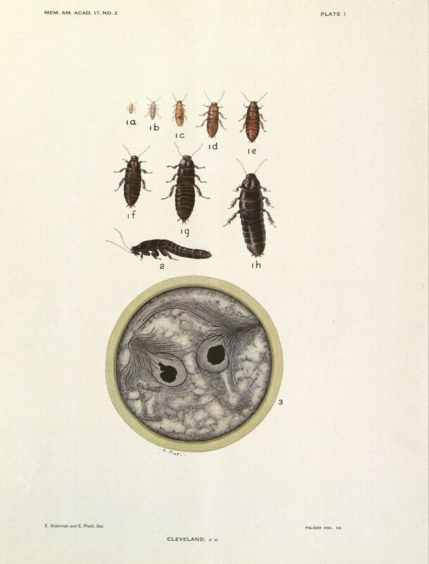
Among these, the oldest is the Kirby-Cleveland collection, which details the microbes that live in the guts of termites and make it possible for these wood-eating insects to thrive on their specialized diet. Assembled beginning in the 1920s by entomologist Harold Kirby and curated by a number of his colleagues across the country following his death, the collection arrived at its permanent home in the Museum in 1991. In 2000, these specimens were joined by a similar collection assembled by late Harvard entomologist L.R. Cleveland.
The Kirby-Cleveland collections consists of thousands of specimens on slides, as well as photographs, illustrations, and even blown-glass models of gut bacteria. These specimens helped propel the modern study of symbiotic relationships and are still consulted by researchers today.
“This collection presages the founding of the field of symbiosis. Before the ability to sequence DNA, researchers would pick up a termite, squeeze out its poop, and find a lot of microscopic life, like protists and bacteria, that let it feed on just wood,” says Susan Perkins, associate curator in the Museum’s Division of Invertebrate Zoology.
Betsey Dyer, a professor of biology at Wheaton College who has worked in the collection, agrees. “The Kirby collection is one of the most complete collections… of a morphologically complex microbial group that diversified into thousands of species,” Dyer says.
Kirby’s collecting also led to the discovery that geography can shape the makeup of a termite’s gut population, Perkins says, pointing out that termites from Africa have distinctly different microbial membership than those in America.
The Museum also has a living collection of single-celled algae and hosts active research on their surprisingly complex biology. In 2012, Assistant Curator Eunsoo Kim showed that some algae don’t live by photosynthesis alone—they also hunger for an occasional bite of bacteria.
New information about microbes isn’t just present in collections devoted to them, though. Perkins points out that, for instance, the Museum’s growing Ambrose Monell Cryo Collection (AMCC) of frozen tissue samples holds more than meets the eye.
“Because we have these tissue samples, we also have the microorganisms that live in that tissue,” says Perkins, who has harvested the malaria-causing parasites she researchers from the frozen tissue of bats and lizards in the AMCC. “We have specific microbe collections, but really, the entire collection is available for study.”