Bats
 Alvaro Keding/© AMNH
An array of bat specimens—including 28 skulls, 45 study skins, an articulated skeleton, and a taxidermy mount—is on display on the third floor of the Louis V. Gerstner, Jr. Collections Core in the Museum's Richard Gilder Center for Science, Education, and Innovation.
The display is an excellent example of how distinct preparation methods can tell you very different things about an animal. The array, along with the variety of species presented, highlights the work of Curator Nancy Simmons, Department of Mammalogy, who studies the evolution and biodiversity of bats.

Skulls and Skeletons
Aside from the obvious size variation among bat species, there is also a wide diversity in the shape of their skulls. These differences in form relate to the diet of the bat (snout length) and to their behavior. Most bat species emit high frequency sound pulses and listen for their echo. This behavior, echolocation, allows the animal to identify the distance, shape, and size of objects in their environment.
 Bat skull, AMNH 107924 Cynopterus titthaecheilus titthaecheilus
Janet Spiller/© AMNH
 AMNH 51738 Hipposideros armiger armiger
Janet Spiller/© AMNH
This exhibit showcases 28 small bat skulls that, for the most part, required limited conservation treatment. Sixteen specimens that had been stored in glass jars were cleaned with a soft brush and then transferred to new vials for display. The others, which would be displayed without containers, required some modest interventions to mitigate the risks associated with their increased exposure. For these, a reversible adhesive was used to stabilize teeth and secure points of contact between articulated bones.

A fully articulated skeleton was among the group selected for display. Bats have a unique skeletal structure that is adapted for flight with elongated fingers, flattened ribs, and backward-facing legs. The elongated fingers, for instance, create a frame over which the skin of the wings is stretched, while flattened ribs allow the bat to glide in the air.
Each bat specimen has a number that identifies and associates it with scientific records containing data regarding its taxonomic identification, sex, when and where it was collected, and by whom. On skulls and skeletons, this number is often written directly on the bone in archival ink. Specimens are also accompanied by paper labels that usually contain more detail. To preserve their scientific value, it is vital that specimens not become disassociated from these labels through loss or deterioration. In preparation for display, conservators transcribed and documented labels carefully and, when necessary, conducted simple stabilization and repairs.
Skins and Taxidermy
A group of 45 study skins and 1 taxidermy mount showcase the diversity of nasal forms, uropatagium (the membrane between the upper legs of the bat), tail morphology, ears, tragi (the thick cartilage projections of the outer ear), and fur coloration in bats. These are just a few of the anatomical markers used in the field to identify each species.
After collection, these specimens were prepared to preserve the skin as well as membranes and bones in the wings and feet. The study skins are supported by a simple pad of cotton wadding that does not attempt to re-create the shape of the animal's body. They are distinct from taxidermy, in which the preserved skin is supported by a carefully modeled internal form (mannikin) with additional components (e.g., glass eyes) added for the purpose of creating a highly lifelike representation.
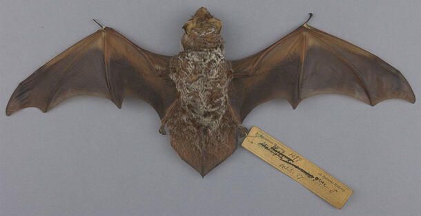
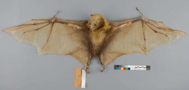
The bat study skins were mostly in good condition and required minimal surface cleaning, as well as some localized linings to support fragile areas on the wings. However the taxidermy Pteropus ocularis specimen arrived at the conservation lab requiring more extensive treatment. Mannikin material inside the mount was visible through an open seam on the ventral side; and significant insect damage and tears were present in the wing membrane (the area of tough skin that extends from the forelimb over the elongated fingers). The tears and small insect holes, along with distortion of the membrane, left the specimen vulnerable to further damage when handled.
The bat was carefully placed face-down on a padded mount, allowing access to the dorsal side of the wings. Goldbeater's skin—a thin, translucent, paper-like material made from animal intestine that was traditionally used as an interleaf between sheets of gold while beating them into gold leaf—was used to repair the skin, filling losses and securing tears.


Nicole Feldman/© AMNH
Using a stable pressure-sensitive adhesive and goldbeater’s skin, repairs were made on the dorsal side of the wings. The adhesive was strong enough to hold the tears together but weak enough to be easily removed. In areas where the goldbeater’s skin was visible, watercolors were used to tone it to blend in with the surrounding membrane.
Pesticides
Historically, animal specimens in many natural history collections were treated with pesticides as a preventive method to deter insects from consuming fur, feathers, and skin. Many of the pesticides used in the past are hazardous to human health and are no longer routinely applied.
X-ray fluorescence spectroscopy (XRF), a non-invasive analytical technique often used to screen for heavy metal pesticide residues in museum collections, detected their presence on the bat study skins. As such, a health and safety protocol was followed in their handling. The protocol included:
- using appropriate signage to designate areas where work with the bats would be completed, and to indicate the presence of heavy metals
- wearing nitrile gloves and lab coats when handling the specimens
- handwashing after handling
- covering lab benches with a paper barrier sheet under specimens to reduce the potential for transfer of heavy metals
- disposing of gloves, barrier sheets, and anything in contact with the study skins in a segregated waste bin
Today, collection care professionals are increasingly prioritizing preventive care as a means to reduce the need for conservation treatment by managing the causes of deterioration directly. In protecting vulnerable animal materials from pests, this shift from a reactive approach toward a more proactive one is reflected in best practices that include maintaining a controlled storage environment with tightly-sealed storage enclosures, pest monitoring, and low-temperature treatment as an alternative to pesticides.