Echinoderms
 Alvaro Keding/© AMNH
This striking array in the Lois V. Gerstner, Jr. Collections Core in the Richard Gilder Center for Science, Education, and Innovation showcases more than 40 dry specimen echinoderms, including burrowing urchins, spiny urchins, shingle urchins, pencil urchins, and sand dollars. It was curated by Estefanía Rodriguez, Curator in the Division of Invertebrate Zoology, and highlights the diversity of these marine invertebrates.
What is an echinoderm?
Echinoderms are marine animals belonging to the class Echinoidea and are characterized by their hard, spiny covering.
Skeleton
Almost every echinoid has a skeleton composed of plates bound together to form a solid structure called a "test." Under high magnification, the structure of the plates is made up of a distinctive meshwork, referred to as the stereom, composed of high-magnesium calcite (calcium carbonate with up to 15% magnesium carbonate). This meshwork structure may be effective in thwarting structural damage by preventing cracks in the plates. Just like humans, the skeletal structure of a sea urchin lies embedded within soft tissue.
Spines
 Sea urchin dry specimens, 2174 Colobocentrotus atratus.
Janet Spiller/© AMNH
 Sea urchin dry specimens, 2059 Plococidaris verticillate.
 Janet Spiller/© AMNH
The spines on a sea urchin are multi-purpose, allowing the animal to move, offer up protection against predators, and avoid structural damage to the test. They are mostly made of calcite, with a small amount of glycoprotein to provide flexibility. Spine shape and size vary among different species. The shingle urchin, for instance, has flattened tesselate shingle-like spines that are shaped to withstand the force of incoming surf in the wave impact zone. The whorl spine urchin, on the other hand, has primary spines that are segmented by small crown-shaped extensions.
Teeth
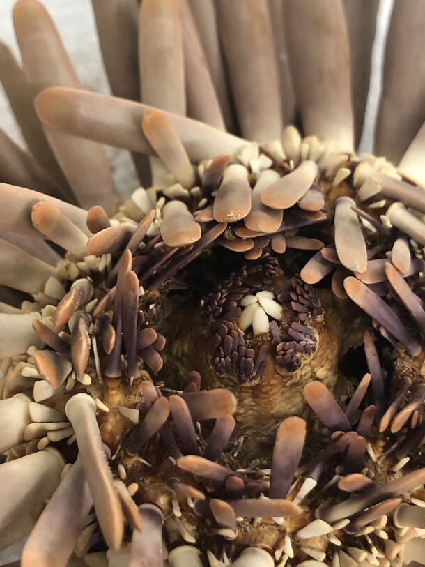
Sea urchins have five sharp teeth set in a circular arrangement that protrude from the center on the oral side of the marine animal. The teeth are very sharp and allow the echinoderm to munch on tough materials, including coral reefs and even rocks. The teeth on a sea urchin are self-sharpening, and researchers are currently using microscopy to study them in hopes of creating new cutting tools. They have found that the teeth are composed of two kinds of calcite crystals, with one side containing fibers that provide a strong supportive brace, while on the other chips away. The calcite is arranged in curved plates with weaker organic material layered in between. Chipping occurs at these weak spots as it scrapes against rock or coral, leaving the edge sharp. The teeth continue growing throughout life, so they never get worn down.
How were the echinoderms conserved for display?
The group of specimens prepared for exhibit in the Collections Core included 31 individual dry sea urchin specimens and 21 sand dollars. Prior to conservation, many of the echinoderms were covered in a layer of fine dust. Additionally, several had detached and/or nearly detached spines. Several examples illustrate these condition issues and treatment approaches for the array.
Slate Pencil Urchin: Disfiguring Dirt
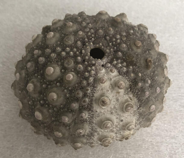
This slate pencil urchin, Heterocentrotus trigonarius, is presented with the test exposed. Its spines were likely lost prior to collection. The aboral side of the urchin (further from the mount) is white with purple pore pairs (pores that support respiratory tube feet). The mamelons (small, rounded ball-like structures on which the spines sit) are exposed, revealing a shiny white surface.
Several of the mamelons had broken edges, revealing matte, gray-purple dirt underneath. There was also thick dirt covering the test and partially covering some of the mamelons. Luckily, the dirt was not tightly bound to the surface, and could be removed using cosmetic sponges and a kneadable cleaning putty (vulcanized cis-polyisoprene rubber) to reveal a whitish/cream colored test
Red Slate Pencil Urchin: Unstable Spines
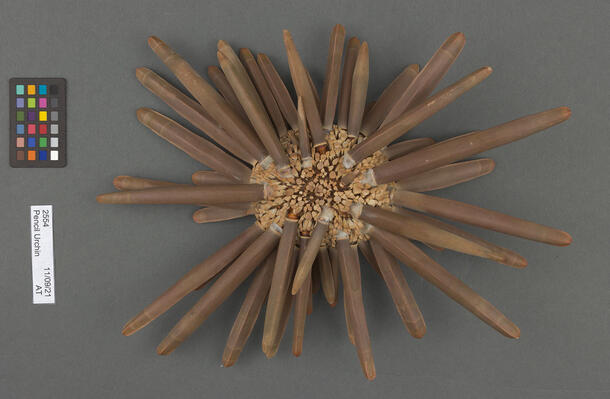
This specimen is a pencil urchin, Heterocentrotus mamilatus, with thick light gray-pink rounded primary spines with two beige rings at the tip. Secondary spines in between the primary ones have a peg-like truncated shape. On the oral side the spines are flatter, smaller, and semi-triangular in cross-section.
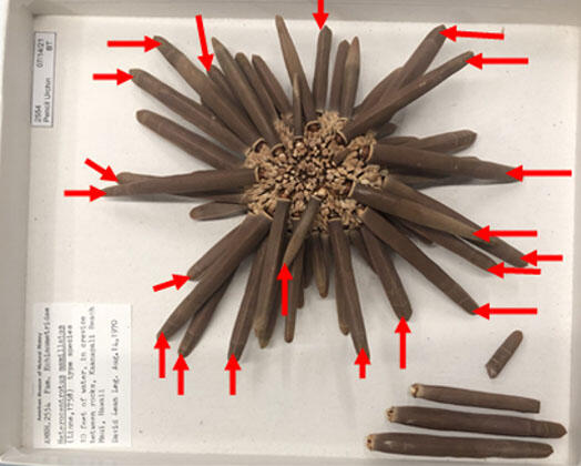
Prior to conservation, the specimen had 23 loose and detached primary spines. One of the detached spines had also broken into two pieces. Many of the loose ones were bent at the base, exposing the mamelons on the test.
After light surface cleaning with cosmetic sponges and cleaning putty, large primary spines were reattached with a reversible, chemically stable acrylic resin adhesive. Loose spines were stabilized by immersing small hand-torn pieces of Japanese tissue paper in the adhesive and carefully placing them over the junction of each spine and mamelon. These "bandages," when dry, are rigid and strong.
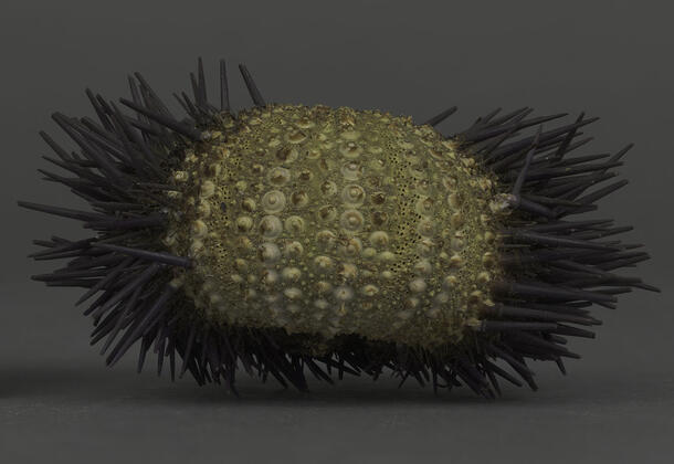
Edible Sea Urchin: Repairing a Broken Test
The last specimen is a hollow (empty) sea urchin, with purple, pink, and dark purple striping on the test. The exposed mamelons on the urchin are small and white.
 Sea urchin dry specimen, 2389A, before treatment, Echinus esculentus.
 Janet Spiller/© AMNH
 Sea urchin dry specimen, 2389A, after treatment, Echinus esculentus.
Janet Spiller/© AMNH
The test was cracked around the periproct (the plated membrane that contains the anus on the aboral side), with two fragments fully detached.
 Sea urchin dry specimen, 2389A, before treatment, Echinus esculentus.
 Janet Spiller/© AMNH
 Sea urchin dry specimen, 2389A, after treatment, Echinus esculentus.
 Janet Spiller/© AMNH
After light dry cleaning, the crack was stabilized with dilute acrylic adhesive applied using a small brush. The join was secured with tape while drying and then reinforced on the interior by lining the crack with a thin strip of Japanese tissue paper. The detached fragments were reattached using the same approach.
Additional Resources
For an in-depth look at echinoderms, including characteristic features of different species and a detailed glossary of terms, see the Natural History Museum of London’s Echinoid Dictionary.