Horns and Antlers
 Alvaro Keding/© AMNH
The horns and antlers array on display on the first floor of the Louis V. Gerstner, Jr. Collections Core in the Richard Gilder Center for Science, Education, and Innovation highlights the natural variety of cranial appendages found in ungulates (hoofed mammals), including giraffids (giraffes and okapis), cervids (deer, caribou and elk) and bovids (antelope, buffalo, sheep, goats, and cattle).
They range in size, color, and character, and have several important functions for animals in life: defense against predators, and demonstration of dominance in mating contexts and control of territory.
Headgear: Ossicones, horns, pronghorns, and antlers
A variety of "headgear” is on display, including ossicones on a giraffe skull, horns on a bison skull, antlers on a deer skull, and pronghorns on an antelope skull. What are these cranial appendages? How are they formed?
Ossicones
Ossicones are found in male okapis and giraffes of both sexes. Female giraffes have two ossicones, while males have a third on the frontal bone. These round, columnar bone protrusions underneath the skin develop initially as cartilage growth in the subcutaneous connective tissue. Over time they harden and fuse to the skull. Unlike horns, they do not protrude beyond the skin. Ossicones contain blood vessels and nerves that are thought to play a role in regulating the temperature of the brain.
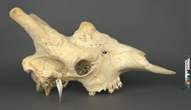
Horns
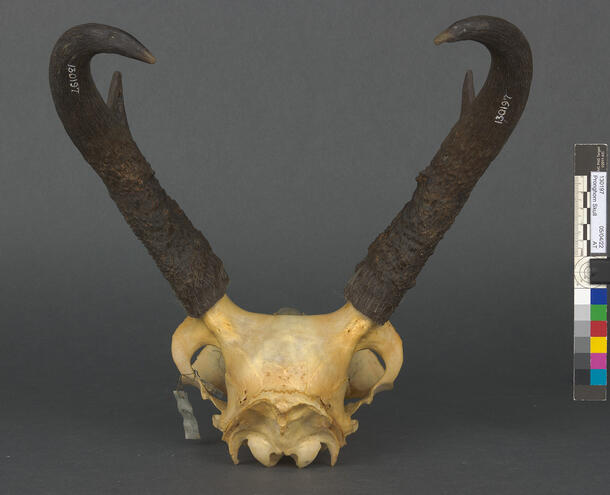
Horns are found on all male bovids, and many females. These permanent projections from the top of the head comprise a boney core and a sheath of keratin, the same material found in feathers and fur. (Read more about keratin in a separate project researching the preservation of feathers and fur).
Horn sheaths can be smooth, curved, ridged, or even fluted. Unlike antlers, horns are not branched, and they grow throughout the entire life of an animal. Horns develop first under the skin as ossicones. Though they are not initially attached to the skull, they later fuse to the frontal bone. Keratin formed in the epidermis covering the bony protrusion is gradually compacted into a specialized sheath. In some species, such as bighorn sheep, the thickness of the compacted keratin varies throughout the year, forming growth rings. These seasonal rings may be related to periods of stress and/or mating behavior.
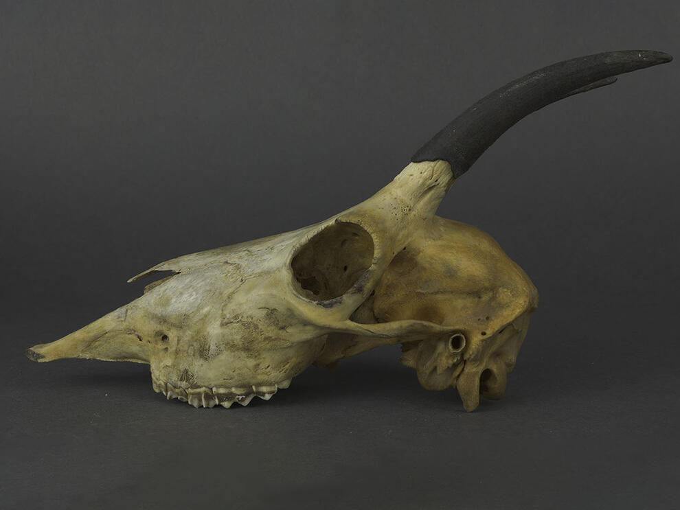
Antlers
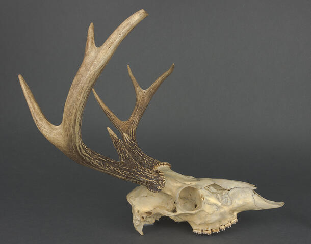
Antlers are found in cervids. With the exception of female caribous, only males have them. In the spring, antlers begin to emerge from pedicles, bony outgrowths of the frontal bone on the animal’s skull. Initially, the antler is covered by fuzzy, vascular skin called velvet that provides nutrition to the fast-growing underlying cartilage and bone. Damage to the pedicle or the velvet can result in abnormal antler growth. Once the antler is fully developed, the velvet is shed and the bone dies. Come winter, the mature antler is shed for the season.
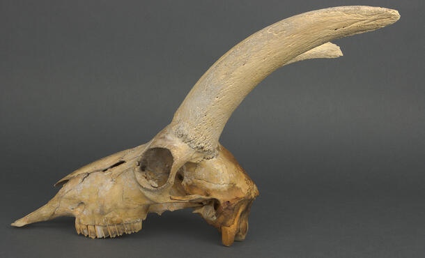
Pronghorns
Pronghorns are structures that share traits with both horns and antlers, but are unique to—as their name suggests—pronghorn antelope (Antilocapra americana). They are found in both the male and female of the species. Pronghorns have a flattened bony core and an overlying keratin sheath. The sheaths however are like antlers in that they branch in males, and are shed annually.
Conservation of skulls
Many of the skulls selected for this array were acquired or donated to the museum in the early 20th century and have remained in storage since. Dirt and grime from years in storage and a yellow patina reveal their age. In a few specimens, some of the sutures that hold together the bones of the skull had separated and become mobile. Many of the teeth were also loose. Their cleaning and consultation was conducted in consultation with Curator Nancy Simmons and Neil Duncan, collections manager in the Museum's Department of Mammalogy.
Adipocere and fatty staining
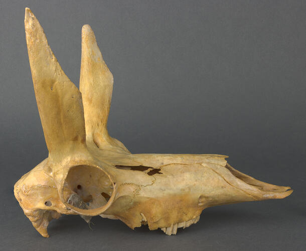
 Mountain goat skull (90294, Oreamnos americanus) with fatty staining, in profile.
Janet Spiller/© AMNH
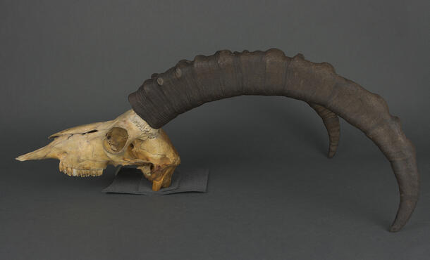
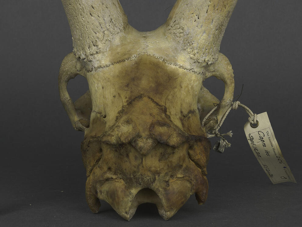 Ibex skull (21476, Capra ibex, without sheaths) with fatty staining, anterior view.
Janet Spiller/© AMNH
A few of the specimens had prominent orange staining on the bone. These areas were tacky and somewhat translucent. Such stains result when natural fats in the specimen are not fully removed through degreasing in preparation. As they oxidize over time, they darken and discolor. This goat skull shows bright orange fatty staining on the rear bones of the skull (occipital, parietal, and temporal bones).
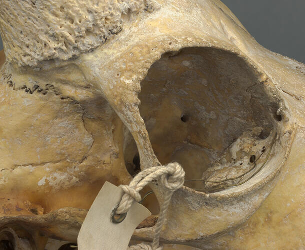
Residual fats also contribute to the formation of adipocere, also sometimes known as corpse wax, coffin wax, mortuary wax or grave wax. Adipocere can appear as a waxy, crumbly, or powdery gray-white or beige accretion on the bone. It is formed through the saponification of fats in the body. In essence, the fat is broken down through anaerobic hydrolysis in an alkaline environment. In this ibex skull, the adipocere is visible as a tenacious grayish-white, sometimes beige, material in and around the eye socket.
Treatment
The skull were cleaned using a variety of dry and wet methods to reduce dirt, grime, adipocere, and fatty staining. Loose teeth were adhered with a stable reversible adhesive and small pieces of Japanese tissue to reduce the likelihood of loss or breakage. After treatment, the skulls were ready to be mounted and installed in the new exhibit.
Time-lapse videos below show examples of the cleaning and stabilization conducted on skulls.