Spiders
Tarantulas
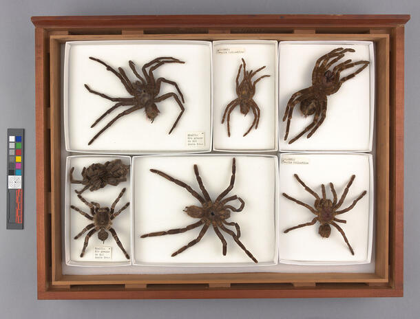
An array of 24 dry tarantula taxidermy specimens can be found on the third floor of the Collections Core display in the Richard Gilder Center for Science, Education, and Innovation. The tarantulas were contributed to the Collections Core by the AMNH Division of Invertebrate Zoology (IZ), and this dynamic display offers a behind-the-scenes glimpse of the storage conditions of an active research collection at AMNH, as if it were a window cut into an IZ storage cabinet.
What is a tarantula?
“Tarantula” is the common name for the generally large, hairy spiders of the Theraphosidae family. There are over 900 described Theraphosidae species, most of which are found in tropical, subtropical, and arid regions of the world. The tarantula specimens in the Collections Core were collected from localities in Brazil, Colombia, and Haiti.
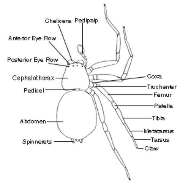
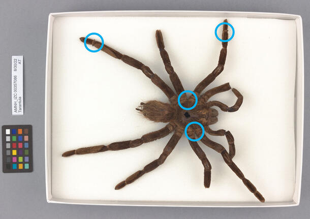
Like all spiders, a tarantula has two main body parts, or tagmata: an abdomen with silk-producing organs called spinnerets, and a fused head and thorax, called the cephalothorax, that bears eight eyes, eight legs, and mouthparts, which include the sensory pedipalps and fanged chelicerae.
Unlike other spider species, tarantulas do not spin webs. Instead, they build silk-lined burrows in which they lay eggs, hide from predators, and lie in wait to ambush prey. Whereas most other spiders have laterally-hinged fangs optimized for seizing prey entangled in their web, tarantulas hunt by pouncing, pinning, and injecting their quarry with two large, downward-facing fangs. Tarantulas typically prey on insects, lizards, and frogs, though some larger species are capable of taking down birds and small mammals for a meal!
A tarantula’s venom is lethal to its small prey, but its bite is relatively harmless to humans and has been described as being about as painful as a bee sting. In fact, tarantulas are quite timid, and their primary defense system is a dense network of “urticating hairs” on their abdomens.
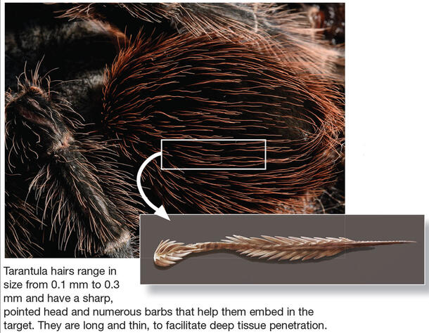
This term is a misnomer, as true hairs are technically only found on mammals; urticating hairs are actually barbed bristles that end in sharp points. When threatened, tarantulas will rub their legs and kick these bristles into the eyes and skin of predators, causing irritation and discomfort. Also unlike true hairs, urticating hairs do not grow back. Instead, a tarantula gains new urticating hairs each time it outgrows and sheds its exoskeleton in a process known as molting.
Strike a pose! How is a dry tarantula specimen prepared?
A tarantula does not have skin; rather, its organs are protected by an exoskeleton composed primarily of chitin. Chitin is a natural polymer found in the exoskeletons of many insects, the shells of crustaceans, and the cell walls of algae and fungi. When a tarantula dies, its exoskeleton dries out and stiffens, and lack of blood pressure in the body causes its legs to fold inward in what is known as the death curl. The preparator therefore must work quickly to remove the organs and replace them with stuffing, and arrange the spider’s limbs to set in the desired pose before they dry.
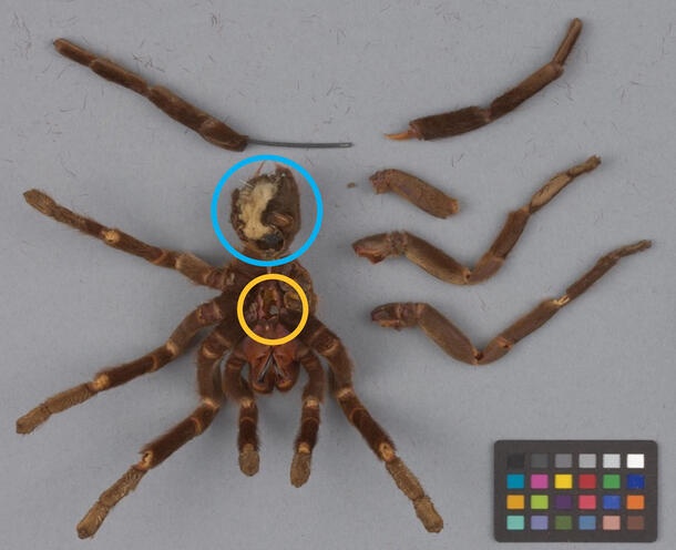
A dry tarantula specimen may be stuffed with cotton to fill the void left by the removal of organs from its abdomen.
The tarantula specimens in the Collections Core were prepared in three distinct poses, each of which informed the conservation treatment methodology:
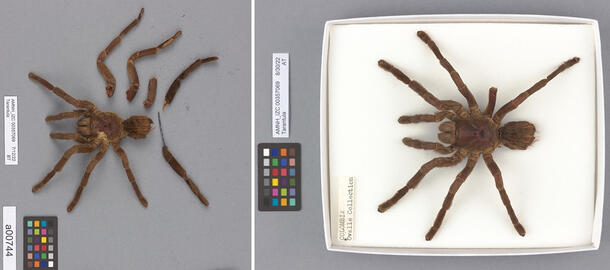
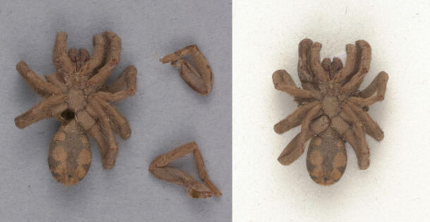
- Spread or splayed
- Standing or walking
- Death curl
How were the tarantula specimens conserved for display?
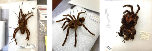
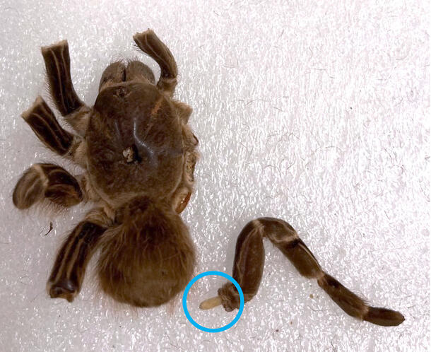
When they arrived in the conservation lab, the tarantulas were in varying condition states. Many of the specimens had fully or partially detached legs, pedipalps, and spinnerets, and in a few cases, the abdomen had completely separated from the cephalothorax. The exoskeletons of all the tarantulas were highly brittle and displayed substantial loss of chitin, especially around the joints of the legs. Additionally, seams from the preparation process had split on many of the tarantulas, revealing the fibrous stuffing materials within their abdomens.
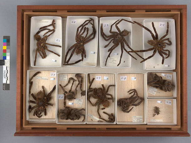
Additionally, each tarantula is associated with at least one paper label containing important data such as the taxonomic identification of the specimen and the date and locality of its collection. These labels were loosely stored with their respective tarantulas in archival trays, many of which were too small for the specimens they held; this placed strain on the fragile joints of the tarantulas, making them vulnerable to breakage and loss. In a research collection, it is crucial that a specimen and its scientific information do not become dissociated.
Dissociation is one of the “agents of deterioration” that threaten cultural heritage collections; you can learn more about the dangers of dissociation and the other agents of deterioration here.
In consultation with AMNH IZ curators, it was decided that the damaged tarantulas should be reassembled for display. The primary treatment goals were (1) to increase visual legibility of the spiders for visitors, and (2) to reduce the risk of loose fragments and paper labels becoming dissociated, which would render them useless in an active research collection.
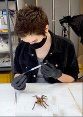
First things first: gloves were worn during treatment to avoid those irritating urticating hairs!
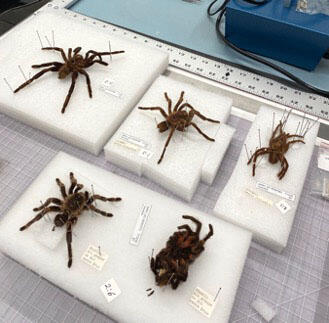
Based on reference photos and anatomical diagrams of tarantulas, the correct placement of each detached fragment was determined. These fragments were gently held in place with insect pins on slabs of foam. The Science Conservation team then developed three distinct approaches to reassembling the fragmented tarantulas that were informed by the size, pose, and stability of each specimen and fragment.
Conservation treatment method 1: Japanese tissue paper dowels
Detached leg and pedipalp fragments could be easily reattached to their respective specimens using small, rolled pieces of Japanese tissue paper that were saturated with a conservation-grade acrylic emulsion adhesive blend and inserted into the hollow appendages and exoskeleton cavities to function like dowels.
The strength and flexibility of these dowels made them the ideal approach for reassembling dry tarantula taxidermy specimens prepared in a walking pose, where the joints are elevated and subject to added strain.
Conservation treatment method 2: direct adhesive application
Detached appendages that were too small or fragile for Japanese tissue paper dowels, such as spinnerets or highly brittle legs, could be directly adhered to the specimen using a syringe loaded with the same acrylic emulsion adhesive blend.
This approach was most effective for the legs of tiny tarantula spiderlings and for larger specimens posed in the death curl, neither of which required the added strength of the Japanese tissue paper dowels.
Conservation treatment method 3
Finally, certain fragments could be arranged and secured with stainless steel insect pins into Ethafoam lining the bottom of each unit tray to give the fragments the appearance of being attached to the tarantula without introducing adhesive to the specimen. This method also did not involve puncturing the specimens, as the round heads of the pins were sufficient to hold the fragments gently and securely in place in a visually subtle manner.

This method was ideal for spiders in the flatter splayed pose. Because it did not involve the use of adhesive, this method was the least interventive, most reversible, and therefore the preferred treatment approach for this project when possible.
Finally, each tarantula was secured with stainless steel insect pins in a new, appropriately-sized paper unit tray lined with foam for support, and each paper label was imaged, transcribed, and pinned into its respective tarantula’s tray, mitigating the risk of dissociation.
The tarantulas were cleaned and reassembled using conservation-informed methods and materials, and the outcome of the treatment is an array of intact, naturalistic, and visually legible research specimens with heightened educational value as representations of their species.
Be on the lookout for our eight-legged friends on display in the Collections Core!