Turtle Shells
 Alvaro Keding/© AMNH
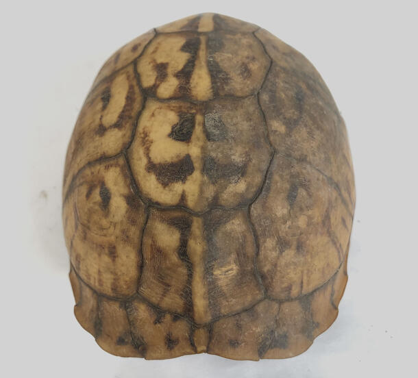
Seventy-six box turtle shells from Gardiner’s Island, with a diversity of coloring and patterning, are on display on the second floor of the Louis V. Gerstner, Jr. Collections Core, part of the Macaulay Family Foundation Collections Galleries, in the Richard Gilder Center for Science, Education, and Innovation. This array highlights the research of Curator Christopher Raxworthy in the Museum's Department of Herpetology.

What is a shell?
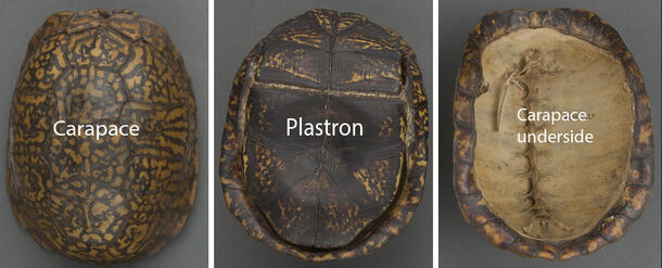
A turtle shell is composed of bone with overlaying scutes. It is formed in two parts: the dorsal portion, or carapace, and the ventral piece, or plastron. The bony shell is both a part of the external protective structure of the turtle, and its internal skeletal structure, with the spine fused to the underside of the carapace.
Covering the exterior sides of the boney carapace and plastron are scutes—hard scales composed of compact keratin. Keratin is the same protein that makes up hair, fingernails, feathers, and horns. The scutes on the box turtle specimens are thin, somewhat flexible, and semi-translucent.
How were the turtle shells originally collected and prepared for the collection?
In conversation with Curator Christopher Raxworthy, it was determined that the shells, already empty with the turtle having died naturally, were collected from Gardiner’s Island, a private island in East Hampton, New York, and brought to the Museum. This particular group of shells received no further preparation as far as the curator knew.
Under different circumstances, similar specimens could be prepared using the Museum’s in-house dermestid beetle colony. The dermestid beetles eat fatty tissue and skin, leaving behind bone and other less desirable parts, such as keratin scutes. While very hungry beetles may eat keratin scutes, they much prefer other tissues. After the fatty remains are eaten away by the colony, the shells can be retrieved.
Goals of Conservation Treatment
Before treatment began, it was necessary to understand goals for the display, and anticipate future research needs. The key questions were:
Q. What narrative about these specimens will be presented to viewers?
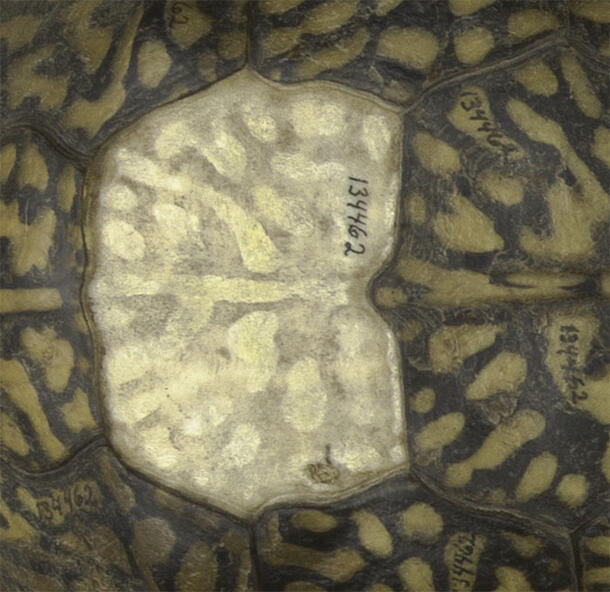
A. The array represents a variety box turtle shells in storage, including various colors and patterns in the scutes, as well as different condition states. Some specimens are missing a few scutes, while almost all the scutes are lost from others. Seven of the box turtle specimens have a partial or full plastron. There are also shells with damaged scutes, green staining, and fatty staining. It was important to highlight various conditions of the shells within this storage context while providing some stabilization for lifting and damaged scutes.
Q. Could cleaning and repair methods affect future DNA analysis or other studies?
A. Applying any solvent or adhesive to bone will inevitably impact analyses conducted in those areas. However, by using solvents minimally and limiting adhesives used for scute stabilization to localized areas, opportunities for future analysis will be preserved. A detailed map of the treatment locations will allow researchers to avoid those areas for testing and sampling.
Q. How should loose scutes be secured to allow for minor movement with environmental fluctuations?
A. The scutes attached to the bone carapace are similar to a medieval polychrome wooden sculpture in that keratin and bone, like paint and wood, expand and contract differently with fluctuations in relative humidity. Over time, as they move relative to one another, the keratin may no longer remain securely attached to the bone and lift or flake off. When choosing an adhesive, we wanted to select something that will allow the scutes some movement while also remaining attached to the shell.
How were the shells conserved for display?
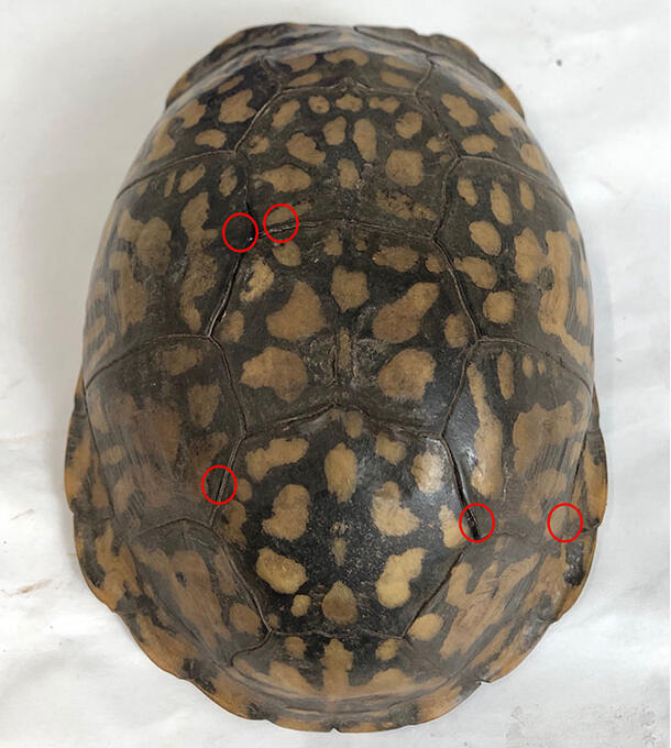
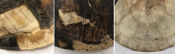
Before treatment began, the turtle shells were examined and their condition described in writing and with photographs. For most specimens, conditions that were commonly noted included lifting scutes and a thick layer of grime and dirt.
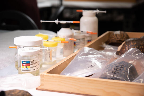
Discreet cleaning and adhesive tests were performed to determine appropriate methods for treatment. Based on the outcome of those tests, a brush and cosmetic sponge were selected for dry cleaning, followed by localized wet cleaning with a mixture of deionized water and ethanol. As current DNA analysis and research are mostly limited to the bony portion of the shells, wet cleaning was confined to the scutes. For some shells, cleaning drastically changed the appearance, strengthening the specimens display value.
A polyvinyl acetate (PVAC) resin was used to resecure detached scutes to the carapace. The adhesive is chemically stable, soluble in many solvents including ethanol and acetone, and has strong adhesion. Additionally, the PVAC has a low glass transition temperature, enabling it to remain flexible as the scutes move over time. To facilitate future DNA sampling, a diagram was created for each shell to map out the location where adhesive was used.
The adhesive took roughly a day to dry, requiring scutes to be held in place during that time.
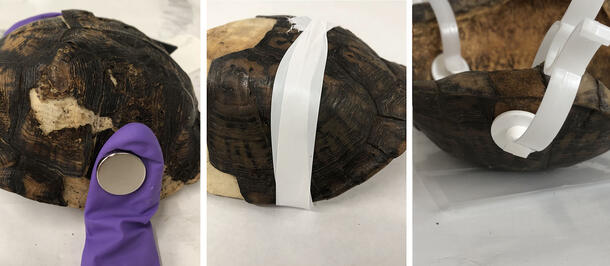
Three methods were successfully used. In the first, small sandbags were made from nitrile glove fingers filled with hydrophobic sand, then secured over the scutes with neodymium magnets. In other cases, PTFE plumber's tape was tightly wrapped around the shell. And when only light pressure was needed, plastic nose clips were effective in reattaching the scutes.