Grouper Skeleton
 Alvaro Keding/© AMNH
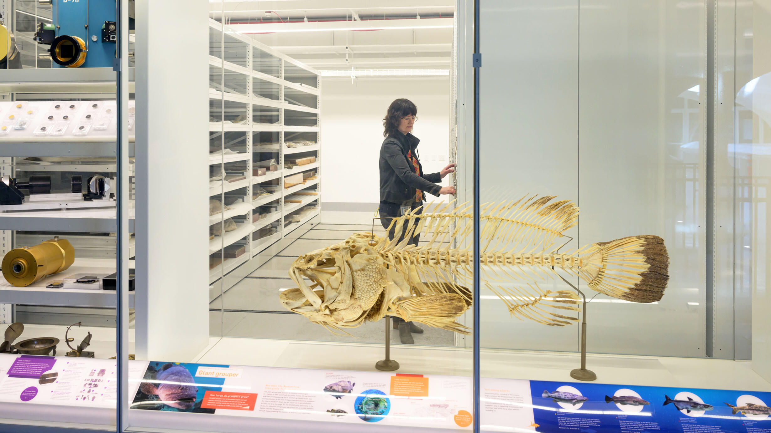
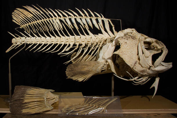
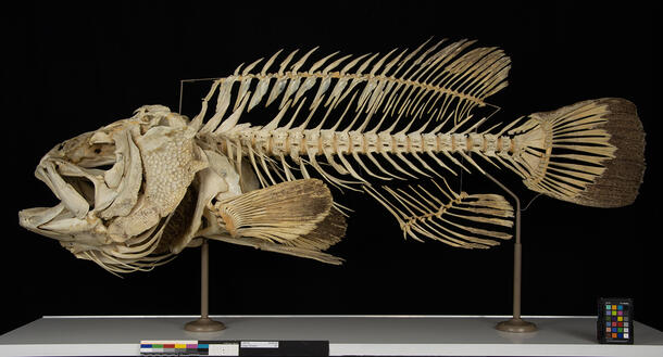
A complete articulated skeleton of the large grouper Epinephelus lanceolatus is among the specimens on display on the third floor of the Louis V. Gerstner Jr. Collections Core in the Richard Gilder Center for Science, Education, and Innovation.
The Museum owns one of the largest collections of fish skeletons in the world, with about 40,000 skeletons. The grouper is a rare specimen, both for its size and because it is one of the only prepared fish specimens in which the skeleton is carefully articulated, with all the bones arranged in the proper order and position.
What are the parts of a grouper skeleton?

The grouper skeleton is composed of scales, bone, gill rakers, and gill filament.
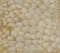
Covering the exterior of the fish are scales, small layered plates that provide armor-like protection. Cycloid scales are preserved on the grouper specimen's operculum. They are composed of two layers: a superficial layer with an organic structure and calcium salts; and a fibrous layer of collagen. Learn more about different types of fish scales.
The majority of the skeleton is composed of bone. Bone is primarily a meshwork of collagen protein with 5-10% calcium phosphate, which imparts its strength and rigidity.
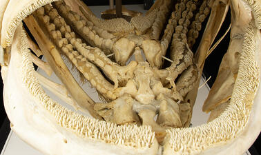
The grouper's gill rakers are tooth-like projections from the gill arch that block debris and small particles from obstructing breathing.
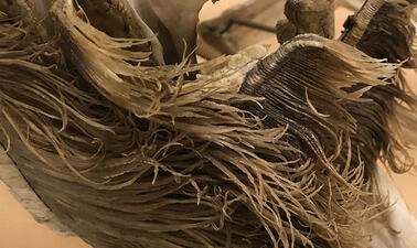
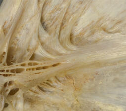
The gill filaments, also called primary lamellae, contain a capillary system that takes up oxygen into the blood, enabling fish to breathe. They are composed on long-chain proteins.
The history of the grouper
The grouper specimen was collected by Curator Gareth Nelson and shipped to the American Museum of Natural History from Darwin, Northern Territory, Australia 25-30 years ago. Archival records indicate that the fish was transported in parts that were individually labeled and prepared using dermestid beetles. Dermestid beetles eat fatty tissue and skin, leaving behind bone and more dense components. While very hungry beetles may eat gill filaments and the collagen layer of the scales, they much prefer other tissues. When a colony of beetles is used to clean a specimen, skeletal components can be retrieved when soft tissue has been removed, but before the beetles damage harder ones.
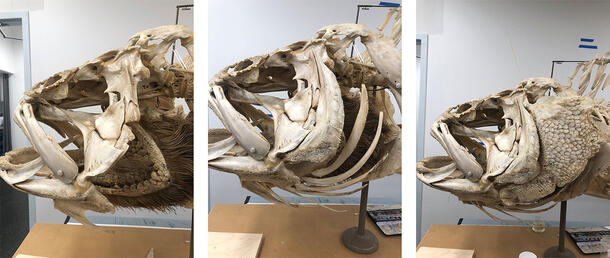
During the skeleton's preparation, the Museum Exhibition team worked with Nelson, who wanted the skeleton to be articulated in a manner that allowed the bones to be easily dissembled for future research and study. The most complicated part of preparing the articulated specimen was construction of the head because it contains many bones that move relative to one another, enabling the fish to open its mouth to feed.
The specimen was placed on display for a short time in the 1990s and then returned to the Department of Ichthyology for storage.
The condition of the grouper skeleton
Throughout the grouper’s time in storage, many bones had become detached from the mount. Large skeletal components and smaller fragments that had become separated include the anal fin ray, caudal fin, left operculum, left preoperculum, and left branchiostegal rays.
In addition, many of the vertebrae, along with a bone on the right branchiostegal ray, were loosely attached. Reconstructed elements, particularly the vertebral spines, were poorly modeled and heavily overpainted. Overall, the surface was dirty with dust, grime, and localized accretions.
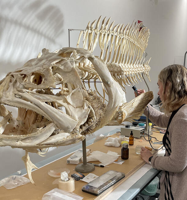
Conservation goals for the grouper
The goals of conservation were defined in consultation with Dean of Science for Collections, Exhibitions, and the Public Understanding of Science, Scott Schaefer, who is the curator-in-charge in the Department of Ichthyology, with input from other curators. The team's aim was to present a realistic, complete skeletal fish specimen and to ensure that the skeleton would remain intact while on display. The effort needed to address structural problems including the re-attachment of detached skeletal elements, as well as aesthetic considerations, including the creation of more realistic intervertebral discs between the vertebrae, and reduction of distracting overpaint.

Reattaching the branchiostegal ray, operculum, and preoperculum
Pre-existing mounting hardware, including hooks and screws, was used to reattach the branchiostegal ray, the preoperculum, and the operculum.
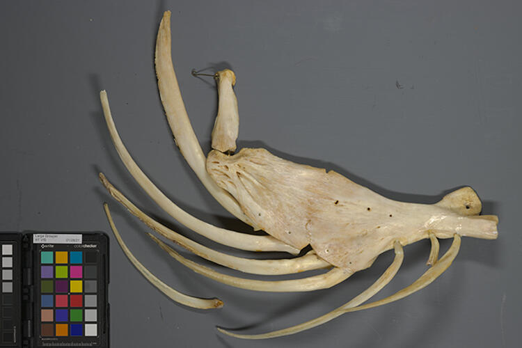 Detached branchiostegal ray.
Nicole Feldman/© AMNH
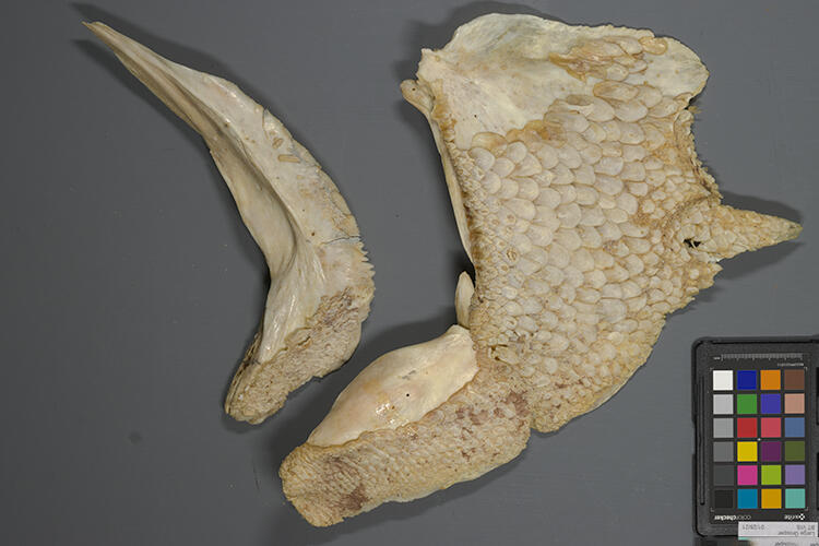 Detached preoperculum and operculum.
 Nicole Feldman/© AMNH
However, where the previous mounting system had failed, stable adhesives and bulking materials were also used to reinforce these attachments. While the specimen was initially prepared in the 1990s so that each bone was easily removable, it was determined in consultation with museum curators that adhesive support, as well as additional points of attachment, were needed. If desired in the future, the adhesive can easily be reversed and the support removed to take apart the bones. In the meantime, they will remain secure while on display.
Reattaching the caudal fin
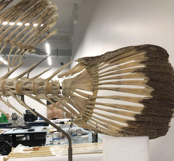
To reattach the tail, a carbon fiber rod was cut to fit inside the caudal fin and the pole supporting the grouper's vertebrae. Carbon fiber is both very strong and extremely lightweight. The join was supported with a bulked adhesive.
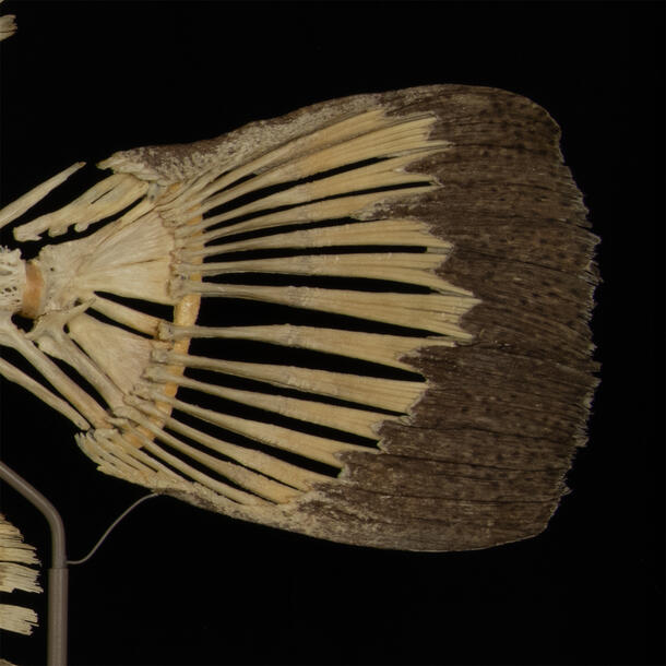
Next, the intervertebral membrane between the last vertebra and the caudal fin was reconstructed, lined with Japanese tissue paper, and painted to provide a realistic appearance. Lastly, a small brass brace was added to provide further support for the caudal fin.
Reattaching the anal fin
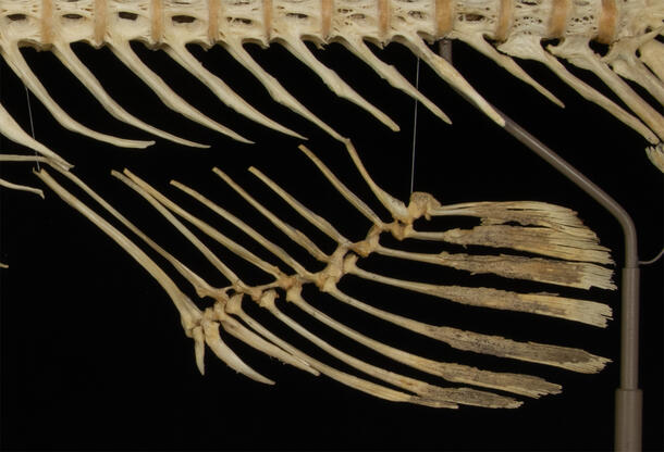
The detached anal fin was reattached using nylon-coated steel beading wire. Tissue was added to disguise the wire and reinforce knots.
Aesthetic considerations
Fill material used between the vertebrae was cracked, and covered with discolored beige paint. After discussions with curators in the Department of Ichthyology, the beige overpaint was removed and the natural look of cartilage between the vertebrae was re-created using the following steps:
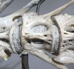
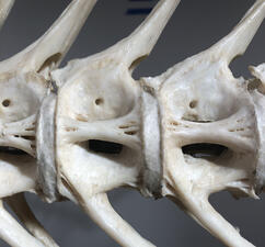
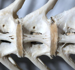
1. Lining the preserved intervertebral membranes with several layers of Japanese tissue paper and adhesive.
2. Applying a toned, translucent heat-set fill material over the tissue paper.
3. Applying a matte acrylic medium to reduce the shininess of the surface.
After cleaning, reattachment of the broken bones, and restoration of the intervertebral membranes, the grouper was ready for display.